

ECG Blog #388 — Why Does Lead V1 Look Funny?
Ken Grauer, MD
JULY 29, 2023
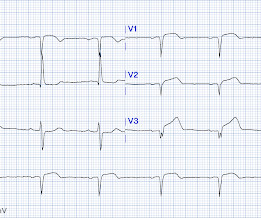
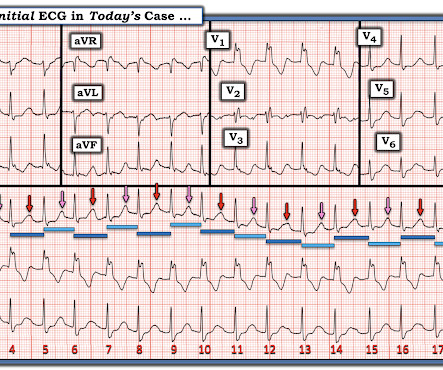
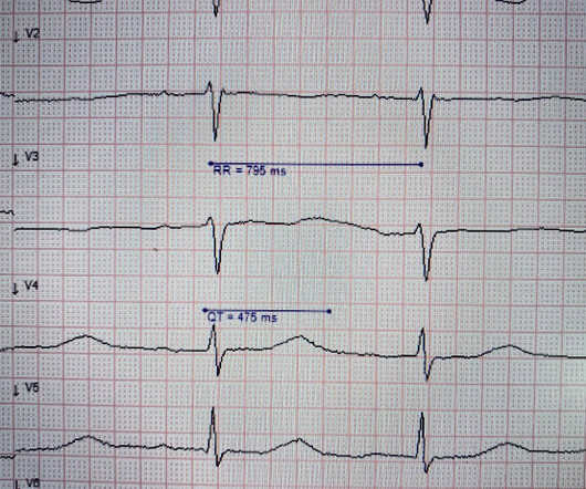
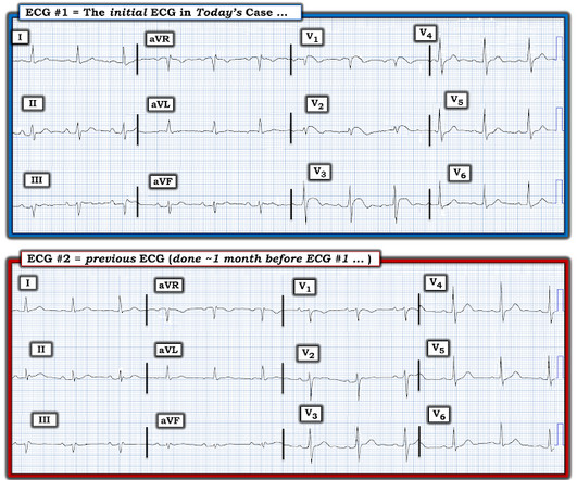
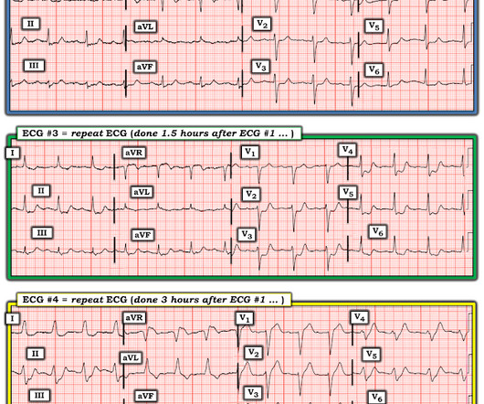
The ECG in Figure-1 was obtained from an 18-year old woman — who moments before been resuscitated from out-of-hospital cardiac arrest. How would YOU interpret her post-resuscitation ECG? Does this ECG in Figure-1 provide clue(s) to the etiology of this patient's cardiac arrest? QUESTIONS: In light of the above clinical history.


Let's personalize your content