article thumbnail

Best Of EAST #5: How Good Is Lung Ultrasound For Pneumothorax?

The Trauma Pro

They advise that the chest ultrasound should be used alone with caution. However, it is at odds with a paper published in 2021 from George Washington University. References: Lung ultrasound underdiagnoses clinically significant pneumothorax. 2021 Nov;49:393-398. false negative rate. EAST 2024, Podium paper 21.

article thumbnail

The Ultrasound-Guided Genicular Nerve Block

ACEP Now

The innervation of the knee is complex, but much of its sensory innervation is supplied by the genicular nerves, which are easy targets for ultrasound-guided nerve blocks. Step 2 Place the ultrasound system contralateral to the affected knee so that you have a clear line of sight of the ultrasound screen when performing the block.

Insiders

Sign Up for our Newsletter

This site is protected by reCAPTCHA and the Google Privacy Policy and Terms of Service apply.

article thumbnail

How Good is Ultrasound at Diagnosing PTA?

RebelEM

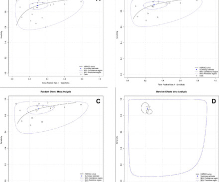
Background: The increased utility and accessibility of point-of-care ultrasound (POCUS) has allowed clinicians the freedom to rethink their diagnostic approach for many common diseases, including peritonsillar abscess (PTA). Test characteristics of ultrasound for the diagnosis of peritonsillar abscess: A systematic review and meta-analysis.

article thumbnail

Trick of the Trade: Gel-free ultrasound-guided peripheral IV technique

ALiEM

You set up everything, have the patient positioned, and then notice there is no sterile ultrasound gel. The trick is to eliminate anything of poor acoustic impedance between the ultrasound probe and the patient’s skin. Note that some ultrasound manufacturers do not recommend the use of isopropyl alcohol on their transducers.

article thumbnail

ICU Physiology in 1000 Words: The Venous Excess Ultrasound Score Is Not the Mean Systemic Pressure

PulmCCM

Kenny MD [ @heart_lung ] Since its first description [1], the Venous Excess Ultrasound Score [VExUS] has received much research attention. Subsequently in the late 1970s, Sivaciyan and Ranganathan [9] confirmed these findings with Doppler ultrasound of the IJV. The ultrasound journal 12: 1-12 2. Jon-Emile S. Echocardiography 7.

article thumbnail

Ultrasound of the Month: Gallbladder Perforation

Taming the SRU

A bedside right upper quadrant ultrasound was performed, and the images are below. The presence of gallstones outside of the gallbladder wall, striated appearance of the gallbladder wall, and adjacent fluid collections, abscesses or fistulas are other findings that could be seen on ultrasound (5, 8). World J Emerg Surg 13, 9 (2018).

article thumbnail

Traumatic Lens Dislocation Identified on Ocular Ultrasound

Cook County EM Blog

On point-of-care ocular ultrasound of the affected eye using the linear probe a round, mobile structure was noted in the posterior chamber with an absence of the lens in its typical position, consistent with posterior lens dislocation. What is the utility of point-of-care ultrasound in identifying lens dislocation? 2021, [link]