article thumbnail

ECG Blog #434 — WHY Did this Patient Arrest?

Ken Grauer, MD

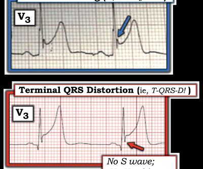
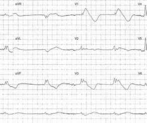
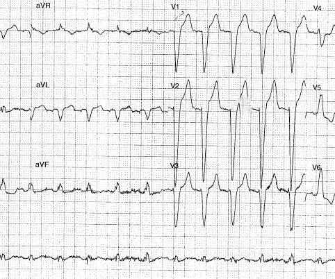
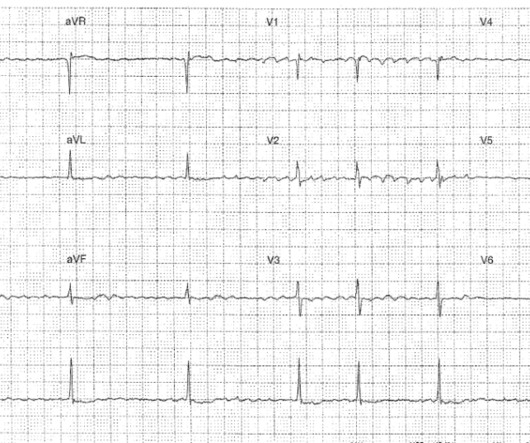
The ECG in Figure-1 — was obtained from a middle-aged man who presented to the ED ( E mergency D epartment ) in cardiac arrest. ROSC ( R eturn O f S pontaneous C irculation ) was obtained — and ECG #1 was recorded. In view of this history — How would YOU interpret the ECG in Figure-1 ? Should you activate the cath lab?

EKG/ECG 399
article thumbnail

ECG Blog #405 — Is AV Block Complete (vs AV Dissociation)

Ken Grauer, MD

For full discussion of this case — See ECG Blog #191 — == The 2-lead rhythm strip shown in Figure-1 was obtained from an elderly woman who presented to the ED following a syncopal episode. ECG Media Pearl # 8 ( 8:20 minutes Video ) — ECG Blog #191 — Distinguishing between A V D issociation vs Complete AV Block ( 2/6/2021 ).

EKG/ECG 432
Insiders

Sign Up for our Newsletter

This site is protected by reCAPTCHA and the Google Privacy Policy and Terms of Service apply.

Trending Sources

article thumbnail

ECG Blog #396 — Why the Flat Line?

Ken Grauer, MD

The ECG in Figure-1 — was obtained from a middle-aged man with palpitations and shortness of breath. How would YOU interpret the ECG in Figure-1 ? Figure-1: The initial ECG in today's case. ( To improve visualization — I've digitized the original ECG using PMcardio ). Figure-1: The initial ECG in today's case. (

EKG/ECG 438
article thumbnail

ECG Blog #415 — The Cath showed NO Occlusion!

Ken Grauer, MD

The ECG in Figure-1 was obtained following successful resuscitation. QUESTIONS: In view of the above history — How would YOU interpret the ECG in Figure-1 ? Is this ECG finding present in today’s initial ECG? Figure-1: The initial ECG in today's case — obtained after successful resuscitation from cardiac arrest. (

EKG/ECG 406
article thumbnail

ECG Blog #409 — Every-Other-Beat.

Ken Grauer, MD

The ECG in Figure-1 — was obtained from a patient with palpitations. Figure-1: The initial ECG in today's case. My Interpretation of the ECG in Figure-1: Since the patient is hemodynamically stable — there is time for systematic assessment of the rhythm. Figure-2: I've labeled the initial ECG in today's case. Figure-2 ).

EKG/ECG 434
article thumbnail

ECG Blog #399 — Which Laddergram is Correct?

Ken Grauer, MD

The ECG in Figure-1 was obtained from a woman in her 60s — who was seen in the ED ( E mergency D epartment ) as part of her evaluation for trauma following a motor vehicle accident. Figure-1: The initial ECG in today's case. To do this — I apply the P s, Q s, 3 R Approach ( See ECG Blog #185 — for review of my system ).

EKG/ECG 395
article thumbnail

ECG Blog #394 — Is QRS Morphology Disguised?

Ken Grauer, MD

The ECG in Figure-1 — was obtained from an older man with a history of prior infarction and coronary bypass surgery. Figure-1: The ECG in today's case. ( To improve visualization — I've digitized the original ECG using PMcardio ). There is no ECG indication of chamber enlargement — ST-T wave changes do not look acute.

EKG/ECG 288