Remove 2020 Remove EKG/ECG Remove Wellness
article thumbnail

ECG Blog #451 — Premature Closure.

Ken Grauer, MD

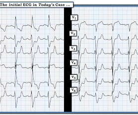
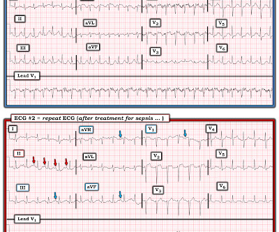
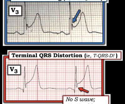
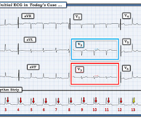
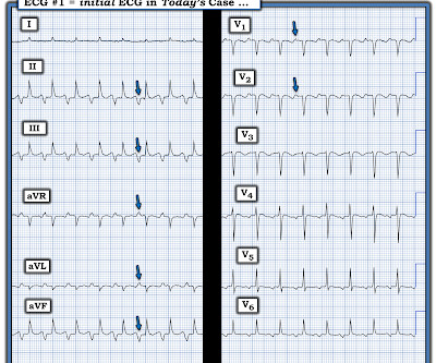
I was sent the ECG shown in Figure-1 — told only that the patient was a middle-aged man with septicemia. Figure-1: The initial ECG in today's case. With practice — it should literally take no more than seconds to assess these 5 Parameters ( See ECG Blog #185 — for more on the Ps,Qs,3R Approach to rhythm interpretation ).

EKG/ECG 425
article thumbnail

ECG Blog #422 — Was Clubbing an ECG Hint?

Ken Grauer, MD

I was sent the ECG in Figure-1 — with the following history: The patient is a young man in his early 20s — who presents to the ED ( E mergency D epartment ) because of SOB ( S hortness O f B reath ) that had been ongoing for several hours. QUESTIONS: In view of the above history — How would YOU interpret the ECG in Figure-1 ?

EKG/ECG 469
Insiders

Sign Up for our Newsletter

This site is protected by reCAPTCHA and the Google Privacy Policy and Terms of Service apply.

article thumbnail

ECG Blog #445 — VT or LBBB?

Ken Grauer, MD

The ECG in Figure-1 was obtained from an 80-year old woman — who presented to the ED ( E mergency D epartment ) — with a several hour history of "palpitations" and CP ( C hest P ain ). She was hemodynamically stable at the time ECG #1 was recorded. Figure-1: The initial ECG in today's case. ( How certain are you of your answer?

EKG/ECG 392
article thumbnail

ECG Blog #406 — To Do Additional Leads?

Ken Grauer, MD

For full discussion of this case — See ECG Blog #351 — == The ECG in Figure-1 — was obtained from a previously healthy older man who contacted EMS ( E mergency M edical S ervices ) because of "chest tightness" that began ~1 hour earlier. Given this history: QUESTIONS: How would YOU interpret the ECG in Figure-1 ?

EKG/ECG 418
article thumbnail

ECG Blog #396 — Why the Flat Line?

Ken Grauer, MD

The ECG in Figure-1 — was obtained from a middle-aged man with palpitations and shortness of breath. How would YOU interpret the ECG in Figure-1 ? Figure-1: The initial ECG in today's case. ( To improve visualization — I've digitized the original ECG using PMcardio ). Figure-1: The initial ECG in today's case. (

EKG/ECG 446
article thumbnail

ECG Blog #430 — Just a Regular LBBB ECG?
Ken Grauer, MD

The ECG in Figure-1 — was obtained from an older man who had just completed dialysis — and , is now complaining of abdominal discomfort that radiates to his chest. Figure-1: The initial ECG in today's case. ( To improve visualization — I've digitized the original ECG using PMcardio ). How would YOU approach this case?

EKG/ECG 405
article thumbnail

ECG Blog #447 — A "Prophetic" P Wave.

Ken Grauer, MD

I was sent the ECG shown in Figure-1 — being told only that providers on the case suspected AFib ( A trial Fib rillation ) with RBBB ( R ight B undle B ranch B lock ) aberrancy. Figure-1: The initial ECG in today's case. ( Figure-1: The initial ECG in today's case. ( How certain are YOU of your answer?

EKG/ECG 248