Remove 2020 Remove EKG/ECG Remove Resuscitation
article thumbnail

ECG Blog #436 — Bigeminy or Alternans?

Ken Grauer, MD

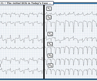
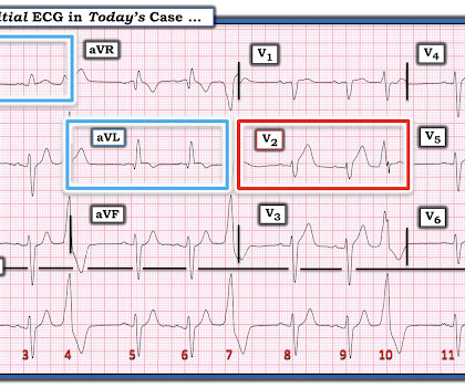
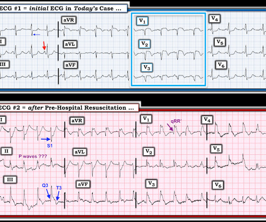
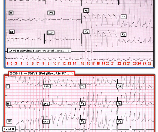
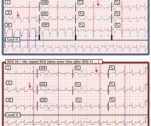
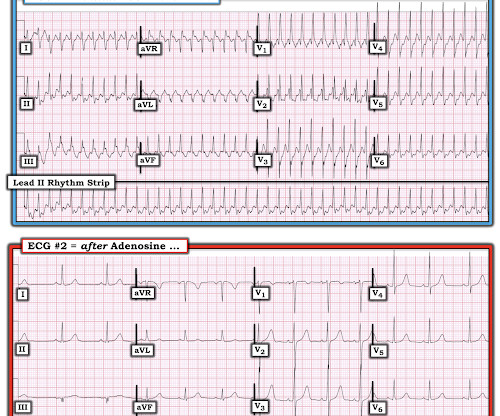
The ECG in Figure-1 — was obtained from an older man with known coronary disease. He developed cardiac arrest shortly after the ECG in Figure-1 was recorded. QUESTIONS: How would YOU interpret the ECG in Figure-1 ? QUESTIONS: How would YOU interpret the ECG in Figure-1 ? Figure-1: The initial ECG in today's case. (

EKG/ECG 400
article thumbnail

ECG Blog #442 — And then the Patient Arrested.

Ken Grauer, MD

I was sent the ECG shown in Figure-1 — told only that that this middle-aged woman noted chest tightness and nausea an hour earlier. QUESTIONS: How would YOU interpret the ECG in Figure-1 ? — IF told that this patient arrested shortly after ECG #1 — Does this change your interpretation? Intervals ( PR-QRS-QTc ) are normal.

EKG/ECG 394
Insiders

Sign Up for our Newsletter

This site is protected by reCAPTCHA and the Google Privacy Policy and Terms of Service apply.

Trending Sources

article thumbnail

ECG Blog #427 — To Cath this Elderly Patient?

Ken Grauer, MD

The ECG in Figure-1 — was obtained on the scene by EMS ( E mergency M edical S ervices ). He was hemodynamically stable — but clearly distressed with a sense of “impending doom” at the time ECG #1 was recorded. QUESTIONS: How would YOU interpret the initial ECG in Figure-1 ? Extra Credit: How many beats are seen on this ECG?

EKG/ECG 338
article thumbnail

A 40-something presented after attempted prehospital resuscitation with persistent Ventricular Fibrillation

Dr. Smith's ECG Blog

A 40-something with persistent Ventricular Fibrillation presented after attempted prehospital resuscitation A 40-something with no previous cardiac history presented to the ED in persistent Ventricular Fibrillation after attempted prehospital resuscitation. Finally, head-up CPR (which was not used here), makes for better resuscitation.

article thumbnail

ECG Blog #381 — Why was the Troponin Normal?

Ken Grauer, MD

The ECG in Figure-1 was obtained from a 60-ish year old man with a history of coronary disease ( including prior CABG ) — who presented to the ED ( E mergency D epartment ) with new CP ( C hest P ain ) of 3 hours duration , diaphoresis and nausea/vomiting. The patient was hypotensive at the time his initial ECG in Figure-1 was obtained.

EKG/ECG 195
article thumbnail

Guidelines would (erroneously) say that this patient who was defibrillated and resuscitated does not need emergent angiography

Dr. Smith's ECG Blog

Here was his initial ED ECG: Formal interpretation by interventional cardiologist: There is "Non-diagnostic" ST Elevation in V2-V4 and aVL. These studies did not address OMI ECG findings!!! So if the patient is awake and has ECG OMI findings , there is no reason to believe that angiography should be withheld. 5% vs. 58%!!

article thumbnail

How a pause can cause cardiac arrest

Dr. Smith's ECG Blog

Below is the presentation ECG. The above ECG shows sinus rhythm. While on telemetry monitoring he suffered cardiac arrest and was resuscitated. What ECG finding may have contributed to (or precipitated) the cardiac arrest? After resuscitation and defibrillation , there were no more episodes of TdP.

EKG/ECG 58