Remove 2020 Remove CPR Remove EKG/ECG
article thumbnail

Hypothermia and drowning

Don't Forget the Bubbles

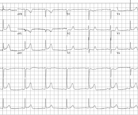
She was intubated at the scene and transported to your ED, with cardiopulmonary resuscitation (CPR) performed en route. You request a 12 lead ECG and repeat a blood gas, asking for it to be run on the PICU analyser. Your trusted nurse hands you the ECG: Paediatric ECG interpretation has never been your strong suit.

article thumbnail

What is the rhythm?

Dr. Smith's ECG Blog

Bystander CPR. Here is the only prehospital 12-lead: Sinus tachycardia, somewhat wide QRS, Ischemia Here is the first ED ECG: What is the rhythm here? This ECG is pathognomonic of hyperkalemia , with wide QRS, very SHARPLY peaked T-waves, flat ST segments, RBBB pattern and large R-wave in aVR. Medics found patient in PEA arrest.

EKG/ECG 109
Insiders

Sign Up for our Newsletter

This site is protected by reCAPTCHA and the Google Privacy Policy and Terms of Service apply.

article thumbnail

A 40-something presented after attempted prehospital resuscitation with persistent Ventricular Fibrillation

Dr. Smith's ECG Blog

Two recent interventions have proven in randomized trials to improve neurologic survival in cardiac arrest: 1) the combination of the ResQPod and the ResQPump (suction device for compression-decompression CPR -- Lancet 2011 ) and 2) Dual Sequential defibrillation. Finally, head-up CPR (which was not used here), makes for better resuscitation.

article thumbnail

1 hour of CPR, then ECMO circulation, then successful defibrillation.

Dr. Smith's ECG Blog

ECMO Flow was achieved after approximately 1 hour of high quality CPR. A followup ECG was recorded 2 days later: No definite evidence of infarction. As per Dr. Smith — “ S hark F in” m orphology was noted on the initial monitoring strip, and initial 12-lead ECG. Figure-1: The initial 12-lead ECG in this case ( See text ).

CPR 52
article thumbnail

Case Report: Coronary Vasospasm-Induced Cardiac Arrest

ACEP Now

During initial assessment, an ECG was obtained and revealed ST-segment elevation (STE) in the inferior leads with ST depression anteriorly. Initial ECG demonstrating inferolateral ST segment elevation and anteroseptal depression, just prior to cardiac arrest. The ECG showed ST-segment elevation without obstructive coronary disease.

article thumbnail

Cardiac Arrest. What does the ECG show? Also see the bizarre Bigeminy.

Dr. Smith's ECG Blog

CPR was started immediately. I was texted this ECG in real time, but it turns out to actually be the 2nd one recorded in the ED. Just as interesting is EKG 1, 24 minutes before the first: What do you think here? Below, I post 4 more examples of ECGs that were recorded from patients who had Torsades. Is this inferior MI?

EKG/ECG 52
article thumbnail

Extreme shock and cardiac arrest in COVID patient

Dr. Smith's ECG Blog

He had the following EKG recorded: Low voltage, suggests effusion. He underwent CPR, and regained a pulse after epinephrine, with an organized narrow complex rhythm at 140, but still with severe shock. This is the ECG on V-V ECMO: Very little STE He had a lot of troponins measured. He was moderately hypoxic.

Shock 52