Remove 2020 Remove COPD Remove Shock
article thumbnail

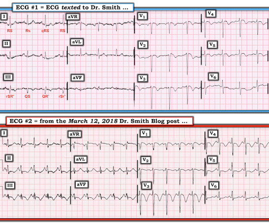
ECG Blog #366 — Diltiazem didn't work.

Ken Grauer, MD

These 2 settings are: i ) In patients with severe , often longstanding pulmonary disease ; and / or , ii ) In acutely ill patients with multi-system disease ( ie, sepsis, shock, electrolyte and/or acid-base disorders ). Applying the Above to Today's Case: In addition to being Covid-positive — the patient in today's case had longstanding COPD.

EKG/ECG 195
article thumbnail

ABG Versus VBG in the Emergency Department

EMDocs

Louis); Marina Boushra, MD (EM-CCM, Cleveland Clinic Foundation); Brit Long, MD (@long_brit) Case Emergency Medical Services brings in a 62-year-old male with COPD in acute on chronic hypoxemic respiratory failure (usually on 3 L nasal cannula, now on non-rebreather at 15 L/min). is more likely to portend death (13, 14). Epub 2019 Dec 30.

Insiders

Sign Up for our Newsletter

This site is protected by reCAPTCHA and the Google Privacy Policy and Terms of Service apply.

Trending Sources

article thumbnail

Diagnostics and Therapeutics: Managing Pneumothorax

Taming the SRU

It can be further divided into two types: primary--those that occur in generally healthy individuals without underlying lung disease, and secondary--those that occur in individuals with underlying lung disease such as COPD [1]. J Emerg Trauma Shock. Published 2020 Jul 23. Published 2020 Jan 21. Ann Emerg Med. Respirology.

article thumbnail

POCUS findings of hemodynamically unstable PE with cardiac arrest

EMDocs

RV chamber size alone is not enough information to rule-in a PE as RV cavity enlargement can be visualized in other conditions such as pulmonary hypertension, RV infarct, COPD and cardiac arrest from multiple causes. 10,11 Vid 1. SubX4 Asystole RV > LV. SubX2 Asystole RV > LV. RV pressures > LV pressures. Tintinalli, J.E.

article thumbnail

A woman in her 20s with syncope

Dr. Smith's ECG Blog

Given her tachycardia and episodes of syncope, the patient was judged to be in compensated obstructive shock with very high risk of imminent decompensation. Figure-2: Causes of Low Voltage on ECG ( Figure reproduced from My Comment at the bottom of the page in the November 12, 2020 post in Dr. Smith's ECG Blog ).

EKG/ECG 52
article thumbnail

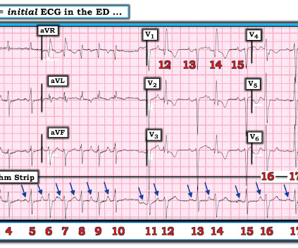
Atrial fibrillation? Multifocal Atrial Tachycardia? Don't look at computer read until AFTER you interpret!

Dr. Smith's ECG Blog

This 60-something with h/o COPD and HFrEF (EF 25%) presented with SOB and chest pain. of all cases, and 62% of Veritas® misdiagnoses). == MY Comment , by K EN G RAUER, MD ( 1/5/2020 ): == This case illustrates a number of important teaching points. Here is the ECG: What do you think? Computer interpretation is below.

EKG/ECG 52
article thumbnail

Are these Wellens' waves?

Dr. Smith's ECG Blog

This clinical information followed: "The patient had a COPD exacerbation with a prehospital SpO2 of 60%. Tachycardia is unusual in ACS unless there is cardiogenic shock or a second simultaneous pathology. Many patients with acute COPD exacerbations present with a similar heart rate. This is NOT Wellens. Is the patient hypoxic?

EKG/ECG 52