Remove 2020 Remove COPD Remove Resuscitation
article thumbnail

Case Report: Coronary Vasospasm-Induced Cardiac Arrest

ACEP Now

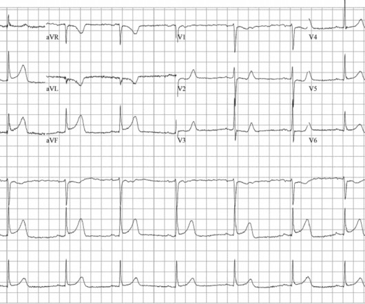
A 45-year-old male with a history of chronic obstructive pulmonary disease (COPD), asthma, amphetamine and tetrahydrocannabinol (THC) use, and coronary vasospasm presented to triage with chest pain. We present a case of refractory ventricular fibrillation resuscitation due to coronary vasospasm from recent amphetamine use with IV NTG.

article thumbnail

Diagnostics and Therapeutics: Managing Pneumothorax

Taming the SRU

It can be further divided into two types: primary--those that occur in generally healthy individuals without underlying lung disease, and secondary--those that occur in individuals with underlying lung disease such as COPD [1]. Unsuspected tension pneumothorax as a hidden cause of unsuccessful resuscitation. Published 2020 Jul 23.

Insiders

Sign Up for our Newsletter

This site is protected by reCAPTCHA and the Google Privacy Policy and Terms of Service apply.

article thumbnail

POCUS findings of hemodynamically unstable PE with cardiac arrest

EMDocs

RV chamber size alone is not enough information to rule-in a PE as RV cavity enlargement can be visualized in other conditions such as pulmonary hypertension, RV infarct, COPD and cardiac arrest from multiple causes. 10,11 Vid 1. SubX4 Asystole RV > LV. SubX2 Asystole RV > LV. Upon arrival, our 25-year-old patient was in PEA arrest.

article thumbnail

A woman in her 20s with syncope

Dr. Smith's ECG Blog

Figure-2: Causes of Low Voltage on ECG ( Figure reproduced from My Comment at the bottom of the page in the November 12, 2020 post in Dr. Smith's ECG Blog ). Figure reproduced from My Comment at the bottom of the page in the September 7, 2020 post in Dr. Smith's ECG Blog ). What is ELECTRICAL ALTERNANS?

EKG/ECG 40
article thumbnail

ABG Versus VBG in the Emergency Department

EMDocs

Louis); Marina Boushra, MD (EM-CCM, Cleveland Clinic Foundation); Brit Long, MD (@long_brit) Case Emergency Medical Services brings in a 62-year-old male with COPD in acute on chronic hypoxemic respiratory failure (usually on 3 L nasal cannula, now on non-rebreather at 15 L/min).

article thumbnail

Chest pain followed by 6 days of increasing dyspnea -- what happened?

Dr. Smith's ECG Blog

Written by Magnus Nossen, edits by Smith The patient in today's case is an 85-year-old male with a history of COPD and dementia. The only other processes identified that caused this type of postinfarction T wave evolution were cardiopulmonary resuscitation, reinfarction, and very small infarcts. The PR interval is normal.

EKG/ECG 90