Remove 2020 Remove COPD Remove EKG/ECG
article thumbnail

ECG Blog #366 — Diltiazem didn't work.

Ken Grauer, MD

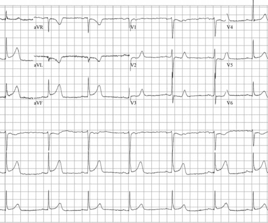
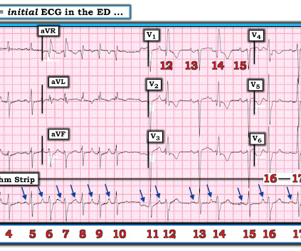
The ECG and long lead II rhythm strip in Figure-1 — was obtained from a COVID positive patient with persistent tachycardia not responding to Diltiazem. Figure-1: The initial ECG — obtained from a patient with persistent tachycardia. ( To improve visualization — I've digitized the original ECG using PMcardio ).

EKG/ECG 195
article thumbnail

VT? Or Supraventricular tachycardic rhythm with aberrancy?

Dr. Smith's ECG Blog

Here is a normal LBBB: In LBBB, monophasic wide R-waves should be limited to the lateral leads in left bundle branch block, as in this ECG. In that ECG above, there are monophasic R-waves starting in lead V2 all the way out to V6. But why does this EKG look nothing like left bundle branch block? Is this sinus tachycardia or VT?

EKG/ECG 133
Insiders

Sign Up for our Newsletter

This site is protected by reCAPTCHA and the Google Privacy Policy and Terms of Service apply.

Trending Sources

article thumbnail

Proportionality is a major element in the ECG Diagnosis of OMI.

Dr. Smith's ECG Blog

This is the result for this ECG, from MDcalc.com : The most accurate cutpoint is 18.2. Here is a similar case from Pendell: This ECG was handed over at triage. That said — the ST-T wave in lead V2 looks to be small only if viewed in the context of its absolute height, as one of 12 leads in the initial ECG shown above in today's post.

EKG/ECG 109
article thumbnail

Case Report: Coronary Vasospasm-Induced Cardiac Arrest

ACEP Now

A 45-year-old male with a history of chronic obstructive pulmonary disease (COPD), asthma, amphetamine and tetrahydrocannabinol (THC) use, and coronary vasospasm presented to triage with chest pain. During initial assessment, an ECG was obtained and revealed ST-segment elevation (STE) in the inferior leads with ST depression anteriorly.

article thumbnail

Regular Wide Complex Tachycardia. What to do?

Dr. Smith's ECG Blog

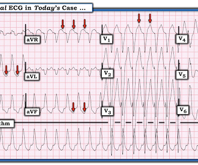
A patient in the ICU with significant underlying cardiac disease [HFrEF 30%, non-ischemic cardiomyopathy, LBBB s/p CRT-D (biventricular pacer), AVNRT s/p ablation a few yrs ago, hx sinus tachycardia while on max tolerated BB therapy] went into a regular wide-complex tachycardia after intubation for severe COPD exacerbation. What do you think?

EKG/ECG 75
article thumbnail

A 40-something woman with acute pulmonary edema -- see the Speckle Tracking echocardiogram.

Dr. Smith's ECG Blog

A 49 year old woman with h/o COPD only presented with sudden dyspnea. Prehospital Conventional algorithm interpretation: ANTERIOR INFARCT, STEMI Transformed ECG by PM Cardio: PM Cardio AI Bot interpretation: OMI with High Confidence What do you think? The ECG findings are focal to the anterior and high lateral wall.

EKG/ECG 52
article thumbnail

Are these Wellens' waves?

Dr. Smith's ECG Blog

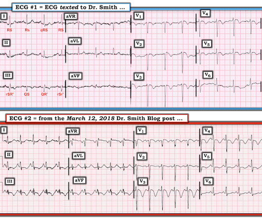
An ECG was texted to me (Smith) without any clinical information: What did I say? This clinical information followed: "The patient had a COPD exacerbation with a prehospital SpO2 of 60%. This clinical information followed: "The patient had a COPD exacerbation with a prehospital SpO2 of 60%. This is NOT Wellens.

EKG/ECG 52