Remove 2020 Remove COPD Remove Documentation/Coding
article thumbnail

Ultrasound in Cardiac Arrest

Mount Sinai EM

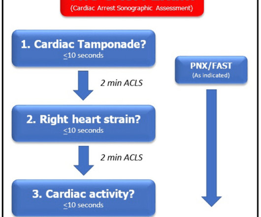
The ideal view depends on the patient’s comorbid conditions such as COPD, obesity, cachexia, etc. Evidence of right heart strain is important but the evidence of fibrinolysis during arrest is mixed with many studies showing no 30-day mortality benefit to lysing during a code. survival to hospital discharge rate.

article thumbnail

ECG Blog #366 — Diltiazem didn't work.

Ken Grauer, MD

Figure-2: I've color-coded P waves from Figure-1 according to P wave morphology ( See text ). NOTE: For clarity — I've color-coded P waves in the long lead II rhythm strip according to morphology. Applying the Above to Today's Case: In addition to being Covid-positive — the patient in today's case had longstanding COPD.

EKG/ECG 195
Insiders

Sign Up for our Newsletter

This site is protected by reCAPTCHA and the Google Privacy Policy and Terms of Service apply.

article thumbnail

Case Report: Coronary Vasospasm-Induced Cardiac Arrest

ACEP Now

A 45-year-old male with a history of chronic obstructive pulmonary disease (COPD), asthma, amphetamine and tetrahydrocannabinol (THC) use, and coronary vasospasm presented to triage with chest pain. Cardiac arrest secondary to myocardial ischemia from coronary vasospasm is well documented. N Engl J Med. 1992;326(22):1451-1455.

article thumbnail

Are these Wellens' waves?

Dr. Smith's ECG Blog

This clinical information followed: "The patient had a COPD exacerbation with a prehospital SpO2 of 60%. Many patients with acute COPD exacerbations present with a similar heart rate. Finally — Note that the S1Q3T3 pattern is missing in ECG #2 , despite documentation of a massive PE. This is NOT Wellens. Is the patient hypoxic?

EKG/ECG 52
article thumbnail

Atrial fibrillation? Multifocal Atrial Tachycardia? Don't look at computer read until AFTER you interpret!

Dr. Smith's ECG Blog

This 60-something with h/o COPD and HFrEF (EF 25%) presented with SOB and chest pain. See below how this has been documented. of all cases, and 62% of Veritas® misdiagnoses). == MY Comment , by K EN G RAUER, MD ( 1/5/2020 ): == This case illustrates a number of important teaching points. Here is the ECG: What do you think?

EKG/ECG 52
article thumbnail

A woman in her 20s with syncope

Dr. Smith's ECG Blog

This patient was reported to have distant heart sounds but was not hypotensive and did not have JVD according to documentation. Figure-2: Causes of Low Voltage on ECG ( Figure reproduced from My Comment at the bottom of the page in the November 12, 2020 post in Dr. Smith's ECG Blog ). What is ELECTRICAL ALTERNANS?

EKG/ECG 40
article thumbnail

ABG Versus VBG in the Emergency Department

EMDocs

Louis); Marina Boushra, MD (EM-CCM, Cleveland Clinic Foundation); Brit Long, MD (@long_brit) Case Emergency Medical Services brings in a 62-year-old male with COPD in acute on chronic hypoxemic respiratory failure (usually on 3 L nasal cannula, now on non-rebreather at 15 L/min).