article thumbnail

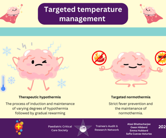
Targeted Temperature Management in Paediatric Traumatic Brain Injury

Don't Forget the Bubbles

His CT scan showed extensive cranial fractures, traumatic subarachnoid haemorrhage, and intraparenchymal haemorrhage. explored the evidence in a 2019 systematic review, which forms part of the Brain Trauma Foundation Guidelines, as did Utsumi et al. 2019 Apr;20(4):404]. At the scene, his lowest GCS was 5 (E1V2M2).

article thumbnail

Neurogenic Shock in Children

Pediatric EM Morsels

Most commonly caused by fracture or dislocation of vertebrae. McGraw Hill Education; 2019:104-109 Dave S, DAHLSTROM JJ. McGraw Hill Education; 2019:125-138 Ziu E, Mesfin FB. Pathophysiology Primary injury happens at the time of the traumatic event or shortly after in the high cervical to mid-thoracic spine. 5 th Edition.

Shock 304
Insiders

Sign Up for our Newsletter

This site is protected by reCAPTCHA and the Google Privacy Policy and Terms of Service apply.

Trending Sources

article thumbnail

Serratus Anterior Plane Blocks for Rib Fractures in the ED

Taming the SRU

Ultrasound‐Guided Serratus Anterior Plane Block (SAPB) Improves Pain Control in Patients With Rib Fractures. This topic is important because of the high incidence of rib fractures in trauma patients and their association with increased morbidity and mortality. Serratus Anterior Plane Blocks for Rib Fractures in the ED.

article thumbnail

Pediatric Hand Fractures

Pediatric Emergency Playbook

Tuft Fracture Yeh PC et al. Pediatric Hand Fractures. Seymour Fracture Nellans et al. Pediatric Hand Fractures. Mallet Fracture Adolescent with mallet finger and Kirschner wire fixation. Pediatric Hand Fractures. Pediatric Hand Fractures. Pediatric Hand Fractures. Pediatric Hand Fractures.

article thumbnail

Proximal Tibiofibular Joint Dislocation in Children

Pediatric EM Morsels

Evaluating the limping child , though, requires us to ponder not only the common (ex, Toddler’s Fracture ), but also to be vigilant for the severe (ex, Septic Arthritis ). Horan, 2006 ] Anatomy of the PTF Joint [ Cunningham, 2019 ] The PTF joint is a synovial joint between the lateral condyle of the tibia and the head of the fibula.

article thumbnail

The Ankle Radiograph

Mount Sinai EM

McGraw Hill; 2019. In: Sherman SC. Simon’s Emergency Orthopedics, 8e. Accessed January 04, 2023. link] Feger J, Knipe H, El-Feky M, Tibiofibular overlap.

Radiology 130
article thumbnail

SGEM#273: My Buddy (Tape) for Boxer’s Fractures

The Skeptics' Guide to EM

Date: October 29th, 2019 Reference: Pellatt et al. Is Buddy Taping as Effective as Plaster Immobilization for Adults With an Uncomplicated Neck of Fifth Metacarpal Fracture? She […] The post SGEM#273: My Buddy (Tape) for Boxer’s Fractures first appeared on The Skeptics Guide to Emergency Medicine. A Randomized Controlled Trial.