Remove 2019 Remove EKG/ECG Remove Ultrasounds
article thumbnail

Torsade in a patient with left bundle branch block: is there a long QT? (And: Left Bundle Pacing).

Dr. Smith's ECG Blog

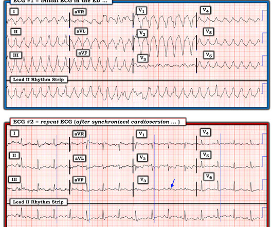
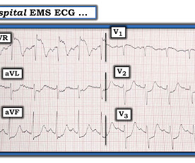
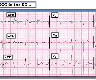
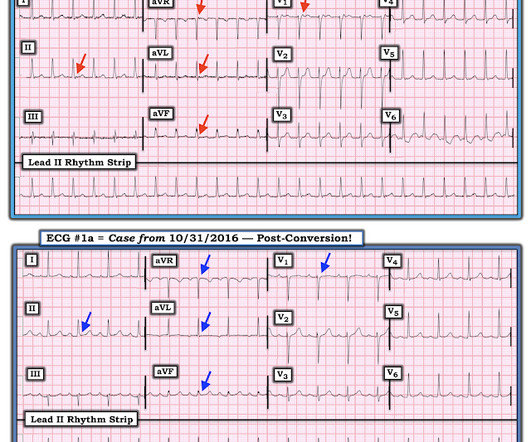
Bedside cardiac ultrasound showed moderately decreased LV function. She had an ECG recorded: This is left bundle branch block (LBBB), with appropriate proportional discordance. Here is one of the strips This is clearly polymorphic VT and probably torsade de pointes Subsequent ECGs. She was intubated. No wall motion abnormality.

EKG/ECG 119
article thumbnail

Four patients with chest pain and ‘normal’ ECG: can you trust the computer interpretation?

Dr. Smith's ECG Blog

All initial ECGs were labeled ‘normal’ or ‘otherwise normal’ by the computer interpretation, and below are the ECGs with the final cardiology interpretation. 1-3] But these studies were very short duration and used cardiology interpretation of ECGs or emergent angiography rather than patient outcomes.

EKG/ECG 122
Insiders

Sign Up for our Newsletter

This site is protected by reCAPTCHA and the Google Privacy Policy and Terms of Service apply.

Trending Sources

article thumbnail

What are treatment options for this rhythm, when all else fails?

Dr. Smith's ECG Blog

The below ECG was recorded. The ECG shows obvious STEMI(+) OMI due to probable proximal LAD occlusion. This ECG does not have the typical ST-vector of an LAD occlusion. See below for Ken Grauer Comment on the initial ECG: == On arrival, another ECG was recorded: There appears to have been quite a bit of spontaneous reperfusion!

EKG/ECG 134
article thumbnail

Noisy, low amplitude ECG in a patient with chest pain

Dr. Smith's ECG Blog

Colin is an emergency medicine resident beginning his critical care fellowship in the summer with a strong interest in the role of ECG in critical care and OMI. An ECG was obtained shortly after arrival: ECG 1 What do you think? The ECG has a lot of artifact, and the amplitude is very small, making interpretation challenging.

EKG/ECG 66
article thumbnail

SGEM#344: We Will…We Will Cath You – But should We After An OHCA Without ST Elevations?

The Skeptics' Guide to EM

The EKG shows sinus tachycardia with nonspecific changes and no ST segment elevations, Q waves, or hyperacute T waves. Her point-of-care ultrasound (POCUS) shows appropriate-appearing global ejection fraction and no marked wall motion abnormalities. The AHA has a statement with recommendations based on the available data.

EKG/ECG 52
article thumbnail

Two patients with chest pain and RBBB: do either have occlusion MI?

Dr. Smith's ECG Blog

Past medical history included RBBB without other cardiac history, but old ECG was not available. As for the ECG, it could represent OMI, but RBBB is also a clue that it may be PE. But with prehospital and ED ECGs being ‘STEMI negative’, the ECG was signed off and the patient waited to be seen.

EKG/ECG 76
article thumbnail

A 20-something with intermittent then acute chest pain

Dr. Smith's ECG Blog

No pericardial effusion on ultrasound." ECG diffuse ST elevation, but lacking pericarditis features, and very concerning for acute injury." It peaked at 21,000 ng/L Post angiogram ECG: The fact that there is near normalization suggests that this may have been a thrombus with complete lysis. What do you think?

EKG/ECG 52