Remove 2019 Remove EKG/ECG Remove Hyperthermia / Hypothermia
article thumbnail

Hypothermia and drowning

Don't Forget the Bubbles

You request a 12 lead ECG and repeat a blood gas, asking for it to be run on the PICU analyser. Your trusted nurse hands you the ECG: Paediatric ECG interpretation has never been your strong suit. What is the likely cause of Elsa’s ECG changes? You look at her monitor, and an arterial blood gas performed moments ago.

article thumbnail

An elderly male with acute altered mental status and huge ST Elevation

Dr. Smith's ECG Blog

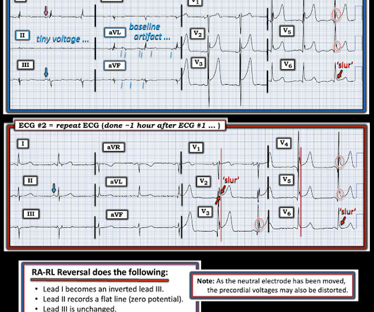
EKG on arrival to the ED is shown below: What do you think? ng/mL (consistent with prior baseline), and a repeat EKG was obtained 1 hour after the initial EKG. limb lead reversal is now resolved) Unfortunately, QOH V1 got tricked by this second ECG! 2) There was no terminal QRS distortion on these ECGs.

EKG/ECG 118
Insiders

Sign Up for our Newsletter

This site is protected by reCAPTCHA and the Google Privacy Policy and Terms of Service apply.

Trending Sources

article thumbnail

A Middle-Aged male with Chest Pain and an Unusual ECG

Dr. Smith's ECG Blog

This ECG was sent from South Asia. Here is the ECG: What do you think? Followup ECG: No Change Absence of evolution is the best evidence against ischemia as the etiology. Could this be a coincidence that the patient was experiencing chest pain and upon ECG capture the atypical brugada pattern was found? Did it subside?

EKG/ECG 52
article thumbnail

The EMERGE Trial: Emergency vs Delayed Catheterization in Survivors of Out-of-Hospital Cardiac Arrest

RebelEM

The TOMAHAWK (Desch 2021), COACT (Abella 2019), and PEARL (Lemkes 2019) trials examined patients with OHCA without ST elevation with a shockable rhythm, and investigators found no difference in delayed versus emergency angiogram. Article: How-Berlemont C, Lamhaut L, Diehl J, et al. Additionally, the trial had several biases.

article thumbnail

A 53 yo woman with cardiogenic shock. Believe me, this is not what you think.

Dr. Smith's ECG Blog

Here was the ECG: There is sinus tachycardia. had at least 2 potential etiologies for developing acute myocarditis: i ) Profound hypothermia ( core temperature = 90°F = 32.2°C This was sent by a reader. A previously healthy 53 yo woman was transferred to a receiving hospital in cardiogenic shock. and K was normal. Our THANKS to Dr.

Shock 52
article thumbnail

Chemical Burns

Mind The Bleep

Circulation Assess heart rate, blood pressure, peripheral and central CRT, pulses and 3 lead ECG. Exposure Expose the patient in a systematic manner while keeping remaining body areas covered e.g. 1 limb at a time, to reduce the risk of hypothermia. 2019 Nov;34(4):276-281. Epub 2019 Nov 29. J Med Toxicol. Acute Crit Care.

Burns 52
article thumbnail

What is this ECG finding? Do you understand it before you hear the clinical context?

Dr. Smith's ECG Blog

Written by Pendell Meyers First try to interpret this ECG with no clinical context: The ECG shows an irregularly irregular rhythm, therefore almost certainly atrial fibrillation. These are Osborn waves usually associated with hypothermia. C), with Cardiac Echo -- A Pathognomonic ECG. Is there a long QT?