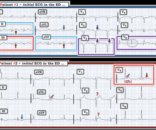
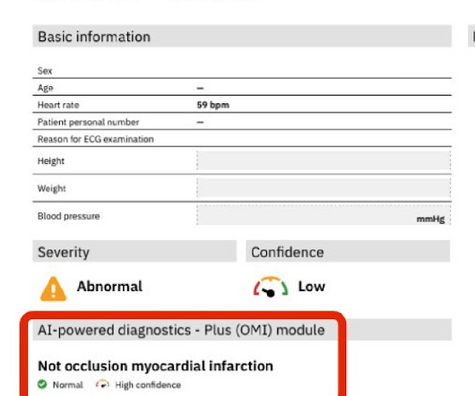
article thumbnail

ECG Blog #451 — Premature Closure.

Ken Grauer, MD

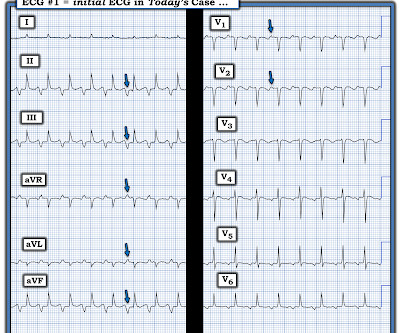
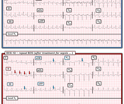
I was sent the ECG shown in Figure-1 — told only that the patient was a middle-aged man with septicemia. Figure-1: The initial ECG in today's case. With practice — it should literally take no more than seconds to assess these 5 Parameters ( See ECG Blog #185 — for more on the Ps,Qs,3R Approach to rhythm interpretation ).

EKG/ECG 425
article thumbnail

ECG Blog #432 — "Should I Shock this Patient?"

Ken Grauer, MD

I was sent the ECG in Figure-1 — without the benefit of any history. Figure-1: I was sent this ECG without any history. MY Thoughts on Today’s CASE: As tempting as it might be to reach for the defibrillator on seeing the ECG shown in Figure-1 — My initial reaction was different. Is this VT? Is this VT? What to do next ?
Shock 414
Insiders

Sign Up for our Newsletter

This site is protected by reCAPTCHA and the Google Privacy Policy and Terms of Service apply.

Trending Sources

article thumbnail

ECG Blog #406 — To Do Additional Leads?

Ken Grauer, MD

For full discussion of this case — See ECG Blog #351 — == The ECG in Figure-1 — was obtained from a previously healthy older man who contacted EMS ( E mergency M edical S ervices ) because of "chest tightness" that began ~1 hour earlier. Given this history: QUESTIONS: How would YOU interpret the ECG in Figure-1 ?

EKG/ECG 418
article thumbnail

ECG Blog #434 — WHY Did this Patient Arrest?

Ken Grauer, MD

The ECG in Figure-1 — was obtained from a middle-aged man who presented to the ED ( E mergency D epartment ) in cardiac arrest. ROSC ( R eturn O f S pontaneous C irculation ) was obtained — and ECG #1 was recorded. In view of this history — How would YOU interpret the ECG in Figure-1 ? Should you activate the cath lab?

EKG/ECG 407
article thumbnail

ECG Blog #415 — The Cath showed NO Occlusion!

Ken Grauer, MD

The ECG in Figure-1 was obtained following successful resuscitation. QUESTIONS: In view of the above history — How would YOU interpret the ECG in Figure-1 ? Is this ECG finding present in today’s initial ECG? Figure-1: The initial ECG in today's case — obtained after successful resuscitation from cardiac arrest. (

EKG/ECG 411
article thumbnail

ECG Video Blog #407 (292): Why the Patient Died?

Ken Grauer, MD

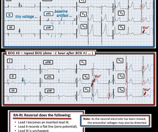
For full discussion of this case — See ECG Blog #292 — == The 2 ECGs shown in Figure-1 were obtained from a man in his 30s — who presented to the ED ( E mergency D epartment ) with chest pain that began several hours earlier. ECG #2 was recorded 1 hour after ECG #1. (22:3 0 min. Initial troponin was negative.

EKG/ECG 412
article thumbnail

ECG Blog #412 — Is Cardiac Cath Indicated?

Ken Grauer, MD

The ECG in Figure-1 was obtained from a middle-aged man with known hypertension — who presented to the ED ( E mergency D epartment ) for CP ( C hest P ain ) over the preceding 2-3 days. QUESTIONS: How would YOU interpret the ECG in Figure-1 ? The Initial ECG in Today’s CASE: The rhythm in ECG #1 is sinus at 65-70/minute.

EKG/ECG 400