Remove 2019 Remove Documentation/Coding Remove EKG/ECG
article thumbnail

ECG Blog #366 — Diltiazem didn't work.

Ken Grauer, MD

The ECG and long lead II rhythm strip in Figure-1 — was obtained from a COVID positive patient with persistent tachycardia not responding to Diltiazem. Figure-1: The initial ECG — obtained from a patient with persistent tachycardia. ( To improve visualization — I've digitized the original ECG using PMcardio ).

EKG/ECG 195
article thumbnail

Death Verification

Mind The Bleep

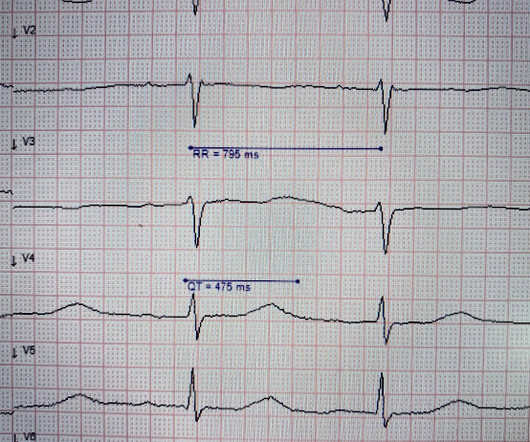
Assessment A code of practice for confirmation of death exists, however, each hospital may have its own protocols which you must familiarise yourself with. If there are family are present, greet them and offer your condolences.

Insiders

Sign Up for our Newsletter

This site is protected by reCAPTCHA and the Google Privacy Policy and Terms of Service apply.

Trending Sources

article thumbnail

Four patients with chest pain and ‘normal’ ECG: can you trust the computer interpretation?

Dr. Smith's ECG Blog

All initial ECGs were labeled ‘normal’ or ‘otherwise normal’ by the computer interpretation, and below are the ECGs with the final cardiology interpretation. 1-3] But these studies were very short duration and used cardiology interpretation of ECGs or emergent angiography rather than patient outcomes.

EKG/ECG 122
article thumbnail

An elderly male with acute altered mental status and huge ST Elevation

Dr. Smith's ECG Blog

EKG on arrival to the ED is shown below: What do you think? The providers documented concern for ST elevation in the precordial and lateral leads as well as a concern for hyperkalemic T waves in the setting of succinylcholine administration. limb lead reversal is now resolved) Unfortunately, QOH V1 got tricked by this second ECG!

EKG/ECG 118
article thumbnail

Torsade in a patient with left bundle branch block: is there a long QT? (And: Left Bundle Pacing).

Dr. Smith's ECG Blog

She had an ECG recorded: This is left bundle branch block (LBBB), with appropriate proportional discordance. In the middle of the night, a "code" was called, and multiple rhythms like this were recorded. Here is one of the strips This is clearly polymorphic VT and probably torsade de pointes Subsequent ECGs. J Am Coll Cardiol.

EKG/ECG 119
article thumbnail

Cardiac Rhythms/ECG Module

Don't Forget the Bubbles

Add into this that the majority of children will be in normal sinus rhythm (NSR) by the time of assessment so to truly identify those who have something wrong we have to be confident in identifying arrhythmias where they are present and critical when analysing an ECG in NSR. All were examined and 98% had an ECG.

EKG/ECG 98
article thumbnail

What are treatment options for this rhythm, when all else fails?

Dr. Smith's ECG Blog

The below ECG was recorded. The ECG shows obvious STEMI(+) OMI due to probable proximal LAD occlusion. This ECG does not have the typical ST-vector of an LAD occlusion. See below for Ken Grauer Comment on the initial ECG: == On arrival, another ECG was recorded: There appears to have been quite a bit of spontaneous reperfusion!

EKG/ECG 134