This site uses cookies to improve your experience. To help us insure we adhere to various privacy regulations, please select your country/region of residence. If you do not select a country, we will assume you are from the United States. Select your Cookie Settings or view our Privacy Policy and Terms of Use.
Cookie Settings
Cookies and similar technologies are used on this website for proper function of the website, for tracking performance analytics and for marketing purposes. We and some of our third-party providers may use cookie data for various purposes. Please review the cookie settings below and choose your preference.
Used for the proper function of the website
Used for monitoring website traffic and interactions
Cookie Settings
Cookies and similar technologies are used on this website for proper function of the website, for tracking performance analytics and for marketing purposes. We and some of our third-party providers may use cookie data for various purposes. Please review the cookie settings below and choose your preference.
Strictly Necessary: Used for the proper function of the website
Performance/Analytics: Used for monitoring website traffic and interactions
Applying the Above to Today's Case: In addition to being Covid-positive — the patient in today's case had longstanding COPD. The September 30, 2019 post in Dr. Smith’s ECG Blog — for an example of “MAT”, but without the tachycardia. He was wheezing, and required supplemental oxygen.
It can be used to distinguish between various conditions, including chronic obstructive pulmonary disease (COPD) exacerbation, acute heart failure (AHF), pleural effusion, pulmonary edema, pericardial effusion, pneumothorax, and pneumonia [2,3]. 2019 Dec 2;102(10):34-38. Published on March 4, 2019. Emerg Med J. PMID: 37595984.
7,10 Contraindications include asthma, chronic obstructive pulmonary disease (COPD), genitourinary or gastrointestinal obstruction, or if there is suspected or confirmed tricyclic antidepressant (TCA) overdose. McGraw Hill; 2019. McGraw-Hill Education; 2019. Physostigmine does not reverse seizures or dysrhythmias.
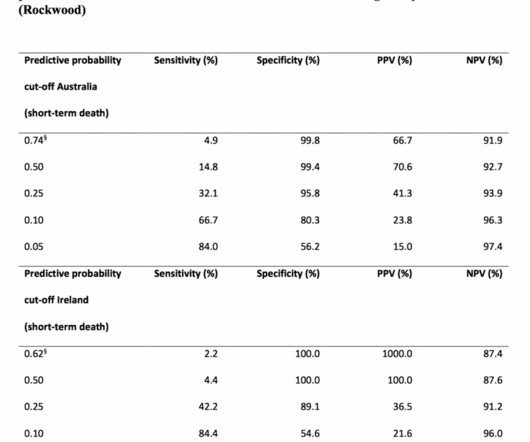
Date: June 28th, 2019 Reference: Cardona et al. AEM June 2019. Date: June 28th, 2019 Reference: Cardona et al. AEM June 2019. AEM June 2019. Prospective Validation of a Checklist to Predict Short-term Death in Older Patients After Emergency Department Admission in Australia and Ireland. Guest Skeptic:Dr.
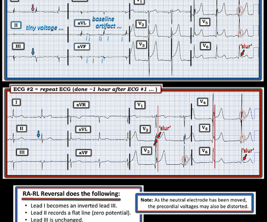
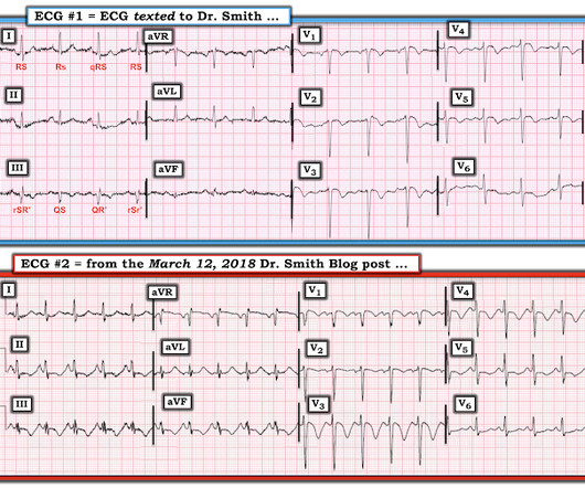
A man in his 90s with a history of HTN, CKD, COPD, and OSA presented to the emergency department after being found unresponsive at home. Rather than loss of both a J wave and S wave — there is a "slur" ( J-point equivalent ) in lead V2 of ECG #2 ( See My Comment in the November 14, 2019 post for illustration of T-QRS-D ).
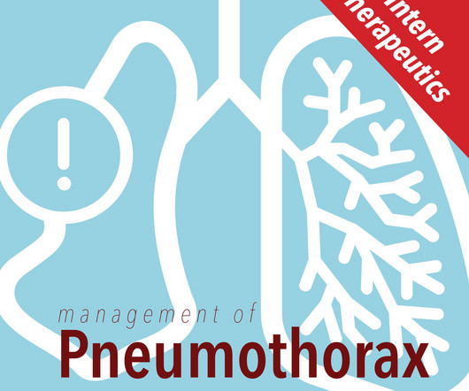
It can be further divided into two types: primary--those that occur in generally healthy individuals without underlying lung disease, and secondary--those that occur in individuals with underlying lung disease such as COPD [1]. Published 2019 Dec 3. doi:10.1155/2019/5271982 Lichtenstein D, Mezière G, Biderman P, Gepner A.
3 days for anoxic encephalopathy to regain pupillary responses after cardiac arrest, 4-7 days for a DNR/DNI patient to receive NIPPV for COPD exacerbation, etc.) They proposed disease-specific intervals for TLTs (e.g., that they invented more or less based on opinion and gestalt (wide flexibility and discretion were also advised).
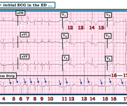
This clinical information followed: "The patient had a COPD exacerbation with a prehospital SpO2 of 60%. These points have been emphasized in many previous posts on Dr. Smith’s blog ( See the April 5, 2019 post , including My Comment at the bottom of that page ). This is NOT Wellens. Is the patient hypoxic? The answer was yes.
This 60-something with h/o COPD and HFrEF (EF 25%) presented with SOB and chest pain. IJC Heart and Vasculature 25(2019). Here is the ECG: What do you think? Computer interpretation is below. Many arrhythmias will prove uninterpretable — IF only 1 or a few leads are used. GET a 12-lead! Providers FORGET to “ U se t he O dds”.
RV chamber size alone is not enough information to rule-in a PE as RV cavity enlargement can be visualized in other conditions such as pulmonary hypertension, RV infarct, COPD and cardiac arrest from multiple causes. 2019 Oct-Dec;29(4):169-171. 10,11 Vid 1. SubX4 Asystole RV > LV. SubX2 Asystole RV > LV. J Cardiovasc Echogr.
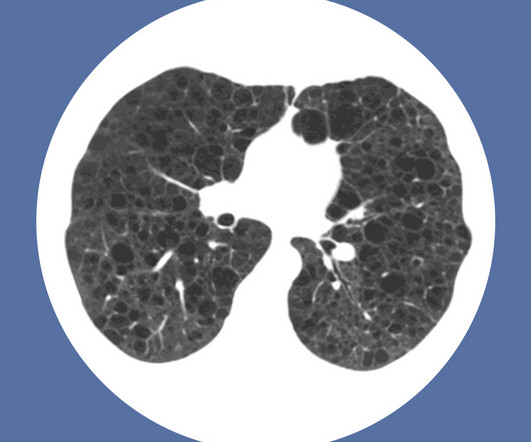
Chronic obstructive pulmonary disease (COPD) is a chronic disease of the lungs caused by inflammatory and structural changes of the small airways and parenchyma of the lungs that result in chronic airflow obstruction and gas trapping. In 2019, the global prevalence of COPD was estimated to be 10.3 Click to enlarge.
Louis); Marina Boushra, MD (EM-CCM, Cleveland Clinic Foundation); Brit Long, MD (@long_brit) Case Emergency Medical Services brings in a 62-year-old male with COPD in acute on chronic hypoxemic respiratory failure (usually on 3 L nasal cannula, now on non-rebreather at 15 L/min). Journal of Trauma and Injury 2019; 32(4): 238-242.
COPD is characterized by episodes of exacerbations, which require prompt recognition and management due to their potential to escalate rapidly and cause significant morbidity and mortality. Epidemiology Globally, more than 300 million people are affected by COPD, marking it as the third leading cause of death worldwide.
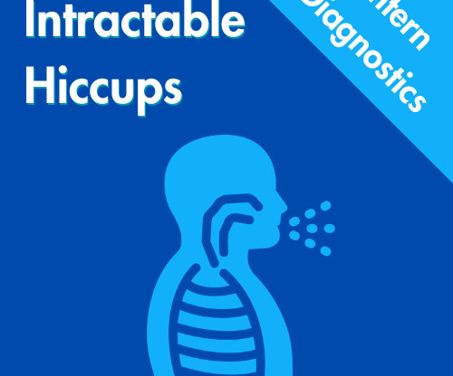
Most common triggers include respiratory conditions such as asthma, COPD, and pneumonia which can lead to forceful coughing -Other common triggers include forceful exertion, severe coughing or vomiting, asthma exacerbation, intense physical activity, or Valsalva maneuvers (e.g., 2019 Jun;217(6):1047-1050. 2019 Oct;45(5):927-931.
Written by Willy Frick A man in his 50s with COPD presented with dizziness and hypotension. As I've emphasized in the February 10, 2025 and March 19, 2019 posts hyperkalemia and hypocalcemia often occur together, and often produce a readily identifiable pattern of flat ST segments ending in peaked T waves that we see in today's case.
Additionally, cardiac instrumentation, recent procedures such as bronchoscopy/endoscopy, central venous catheter placement/displacement, aortic aneurysm, pneumonia, asthma, COPD, pleural effusion, and pericarditis can manifest as intractable hiccups through similar mechanisms. Cham: Springer; 2019. Indian J Psychol Med.
We organize all of the trending information in your field so you don't have to. Join 5,000+ users and stay up to date on the latest articles your peers are reading.
You know about us, now we want to get to know you!
Let's personalize your content
Let's get even more personalized
We recognize your account from another site in our network, please click 'Send Email' below to continue with verifying your account and setting a password.


Let's personalize your content