Remove 2017 Remove EKG/ECG Remove Shock
article thumbnail

Does the literature support medications for stable, monomorphic ventricular tachycardia?

EMDocs

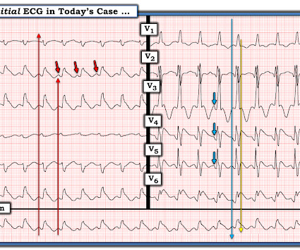
His initial EKG is the following: What do you think? Do we still shock? It was published in European Heart Journal in 2017. If procainamide is utilized, a baseline EKG should be obtained to assess the QRS and QTc at baseline. 2017 May 1;38(17):1329-1335. If the patient has a BP of 60/palp, its easy, right?

EKG/ECG 75
article thumbnail

ACMT Toxicology Visual Pearl: Salt, not Shock

ALiEM

What agent would most likely be responsible for these ECG findings? This EKG shows a wide complex (QRS 240 msec), irregular rhythm with left bundle branch block morphology at a rate slower than expected (90 bpm) for a ventricular arrhythmia such as ventricular tachycardia. 2017 Jul;17(3):260-266. Cardiovasc Toxicol. 2019.158356.

Shock 73
Insiders

Sign Up for our Newsletter

This site is protected by reCAPTCHA and the Google Privacy Policy and Terms of Service apply.

Trending Sources

article thumbnail

Physical Examination as a Helpful Aid in Decision-Making in Challenging ECGs

Dr. Smith's ECG Blog

AslangerE A 65-year-old gentleman presented to the emergency department after experiencing two recent ICD shocks in the preceding hours. An initial electrocardiogram (ECG) is provided below. A subsequent ECG was taken: There is persistent ST elevation, especially being concordant in inferolateral leads. What do you think?

EKG/ECG 112
article thumbnail

VF arrest at home, no memory of chest pain. Angiography non-diagnostic. Does this patient need an ICD? You need all the ECGs to know for sure.

Dr. Smith's ECG Blog

They shocked him twice before return of spontaneous circulation. Here is his presenting ECG: ECG 1, t = 0 What do you think? His transfer packet included notes, labs, cath report, and ECG reports, but no actual ECG images. Smith's ECG Blog. He did not have access to ECG 1.

EKG/ECG 118
article thumbnail

Cardiac Rhythms/ECG Module

Don't Forget the Bubbles

Add into this that the majority of children will be in normal sinus rhythm (NSR) by the time of assessment so to truly identify those who have something wrong we have to be confident in identifying arrhythmias where they are present and critical when analysing an ECG in NSR. All were examined and 98% had an ECG.

EKG/ECG 98
article thumbnail

What are treatment options for this rhythm, when all else fails?

Dr. Smith's ECG Blog

The below ECG was recorded. The ECG shows obvious STEMI(+) OMI due to probable proximal LAD occlusion. This ECG does not have the typical ST-vector of an LAD occlusion. See below for Ken Grauer Comment on the initial ECG: == On arrival, another ECG was recorded: There appears to have been quite a bit of spontaneous reperfusion!

EKG/ECG 131
article thumbnail

Chest discomfort and a dilated right ventricle. What's going on?

Dr. Smith's ECG Blog

The following ECG was recorded. The presenting ECG shows SR with narrow QRS complexes. First troponin I returned 3174 ng/l, at which point a repeat ECG was ordered. Not much difference from 1st ECG. At this point an old ECG on file was found for comparison. Upon admission she had ongoing slight chest discomfort.

EKG/ECG 116