Remove 2017 Remove EKG/ECG Remove Resuscitation
article thumbnail

ECG Blog #388 — Why Does Lead V1 Look Funny?

Ken Grauer, MD

The ECG in Figure-1 was obtained from an 18-year old woman — who moments before been resuscitated from out-of-hospital cardiac arrest. How would YOU interpret her post-resuscitation ECG? Does this ECG in Figure-1 provide clue(s) to the etiology of this patient's cardiac arrest? About A RVC/ A RVD.

EKG/ECG 370
article thumbnail

Does this T wave pattern mean anything?

Dr. Smith's ECG Blog

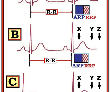
An Initial ECG was performed: Initial ECG: Sinus tachycardia with prolonged QT interval (QTc of 534 ms by Bazett). A repeat ECG was performed 2 hours after arrival: QTc prolongation ato 722 ms now with alternating T wave pattern (T wave alternans) I texted this to Smith who responded: “T wave alternans and long QT. 2005.08.066.

EKG/ECG 131
Insiders

Sign Up for our Newsletter

This site is protected by reCAPTCHA and the Google Privacy Policy and Terms of Service apply.

Trending Sources

article thumbnail

Updates in the Management of Refractory Ventricular Tachycardia or Ventricular Fibrillation Arrest

ACEP Now

17,18 Given this, the advent of extracorporeal membrane oxygenation (ECMO) presents an opportunity to bridge care between traditional resuscitation of refractory VF patients in the ED and more definitive management in the catheterization lab. Resuscitation. Resuscitation. Resuscitation. Resuscitation. N Engl J Med.

article thumbnail

ToxCard: Bupropion

EMDocs

3 Always get an EKG to assess for cardiotoxicity, including widened QRS and prolonged QTc, although this may not be evident right away. 3 Cardiotoxicity 3 All patients need an EKG and cardiac monitoring. Her initial EKG showed sinus tachycardia with a QRS of 86 msec and QTc of 414 msec. Eds (2017). EMIT II immunoassay).

article thumbnail

Electrical injuries

Don't Forget the Bubbles

Suspected low voltage injuries (<1000v) should still be approached with an A-E assessment, but if 12 lead ECG and urinalysis are normal, it’s reasonable to discharge the patient. If the ECG is abnormal, the patient should be managed as if exposed to a high voltage (>1000v). Resuscitation. Culnan, D.M., Jimenez, C.

Burns 80
article thumbnail

Proning for ARDS

Northwestern EM Blog

Hemodynamic instability and ongoing cardiac arrhythmias are a strong contraindication as chest access for cardiopulmonary resuscitation is limited (Koulouras et. Remove ECG leads and patches. Reattach EKG leads to back. 2017 Jan;151(1):215-224. Leave most of the sheet hanging. Turn patient into lateral decubitus position.

article thumbnail

How a pause can cause cardiac arrest

Dr. Smith's ECG Blog

Below is the presentation ECG. The above ECG shows sinus rhythm. While on telemetry monitoring he suffered cardiac arrest and was resuscitated. What ECG finding may have contributed to (or precipitated) the cardiac arrest? After resuscitation and defibrillation , there were no more episodes of TdP.

EKG/ECG 68