Remove 2016 Remove Ultrasounds Remove Wellness
article thumbnail

PEM POCUS Series: Pediatric Lung Ultrasound

ALiEM

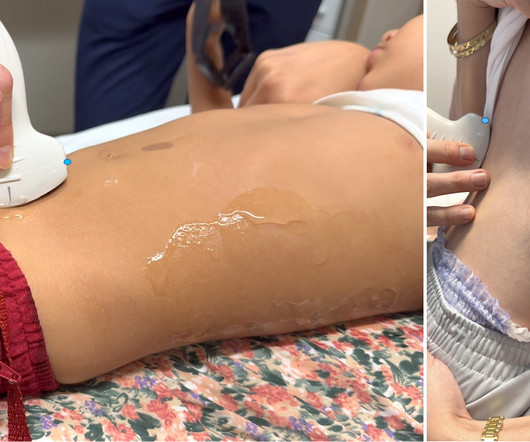
Read this tutorial on the use of point of care ultrasonography (POCUS) for pediatric lung ultrasound. Take the ALiEMU PEM POCUS: Pediatric Lung Ultrasound Quiz Module Goals List indications for performing a pediatric lung point-of-care ultrasound (POCUS). Describe the technique for performing lung POCUS.

article thumbnail

Superior Mesenteric Artery Syndrome (SMA Syndrome) in Children

Pediatric EM Morsels

Oka, 2023) Abdominal ultrasound with doppler can also be used to measure the angle, and with a trained ultra-sonographer was found to be as sensitive as CTA. Positional changes after intake can also be helpful including the lateral decubitus position as well as knee to chest. 2016: 7809281. The Oschner Journal. 15(1):74-78.

Insiders

Sign Up for our Newsletter

This site is protected by reCAPTCHA and the Google Privacy Policy and Terms of Service apply.

Trending Sources

article thumbnail

PEM POCUS Series: Soft Tissue Ultrasound

ALiEM

Take the ALiEMU PEM POCUS: Soft Tissue Quiz Case Goals List the indications of performing a pediatric soft tissue point-of-care ultrasound (POCUS). Pediatric Soft Tissue POCUS Ultrasound Technique Figure 1. Linear ultrasound transducer Probe Use a linear, high-frequency transducer. Describe the limitations of soft tissue POCUS.

article thumbnail

PEM POCUS Series: Pediatric Renal and Bladder Ultrasound

ALiEM

Given her pain with a history of intermittent hematuria and dysuria, you perform a renal and bladder point of care ultrasound (POCUS) examination. Pre-warmed ultrasound gel is helpful when available. Then test your skills on the ALiEMU course page to receive your PEM POCUS badge worth 2 hours of ALiEMU course credit.

article thumbnail

Diagnostics and Therapeutics: Vascular Access in the Emergency Department

Taming the SRU

Ultrasound Guided Peripheral IV (USGIV) Access anatomy of arm veins when considering USGIV. unported license [link] Ultrasound allows us to visualize deeper, typically larger veins in the forearm and upper arm. Place ultrasound machine on the opposite side of the bed to minimize neck strain. Check for flash in the IV chamber.

article thumbnail

Activity Guidelines After Solid Organ Injury: How Important Are They?

The Trauma Pro

years from 2013 to 2016. Clinical outcomes assessed at 60 days included an unplanned return to the emergency department (ED), re-admission, complications, and development of new bleeding confirmed by surgery, ultrasound, or computed tomography (CT) at 60 days post-injury. ” This means they probably did not.

article thumbnail

Acute artery occlusion -- which one?

Dr. Smith's ECG Blog

Answer : Bedside ultrasound! Smith : RV infarct may also have this appearance on ultrasound. So hypoxia without B lines on lung ultrasound strongly weights toward PE. _ Here are some representative stills with red arrows pointing to the emboli. Instead, IR performed pulmonary thrombectomy and the patient did well.

EKG/ECG 128