This site uses cookies to improve your experience. To help us insure we adhere to various privacy regulations, please select your country/region of residence. If you do not select a country, we will assume you are from the United States. Select your Cookie Settings or view our Privacy Policy and Terms of Use.
Cookie Settings
Cookies and similar technologies are used on this website for proper function of the website, for tracking performance analytics and for marketing purposes. We and some of our third-party providers may use cookie data for various purposes. Please review the cookie settings below and choose your preference.
Used for the proper function of the website
Used for monitoring website traffic and interactions
Cookie Settings
Cookies and similar technologies are used on this website for proper function of the website, for tracking performance analytics and for marketing purposes. We and some of our third-party providers may use cookie data for various purposes. Please review the cookie settings below and choose your preference.
Strictly Necessary: Used for the proper function of the website
Performance/Analytics: Used for monitoring website traffic and interactions
Background: The increased utility and accessibility of point-of-care ultrasound (POCUS) has allowed clinicians the freedom to rethink their diagnostic approach for many common diseases, including peritonsillar abscess (PTA). Test characteristics of ultrasound for the diagnosis of peritonsillar abscess: A systematic review and meta-analysis.
We’ll keep it short, while you keep that EM brain sharp. A 74-year-old female with a past medical history of hypertension, diabetes, recent basilar artery stent placement with a 20 pack-year smoking history presents to the ED via EMS for altered mental status and episodes of apnea. Semin Ultrasound CT MR. 2013 Apr;34(2):131-41.
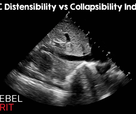
Regarding caval indexes, the advent of artificial intelligence and advanced learning has become integrated into many ultrasound machines. Jan 2015; PMID: 25559473 Airapetian N, et al. Nov 2015; PMID: 26563768 Yartsev, Alex. Ultrasound Med Biol. 3 Cutoff CI of 15% has a negative predictive value (NPV) of 100%.
We’ll keep it short, while you keep that EM brain sharp. 2015 Feb 2;2015(2):CD001067. The post EM@3AM: Endometritis appeared first on emDOCs.net - Emergency Medicine Education. She was discharged 3 days prior after a cesarean delivery to a single, full term, live born infant complicated by premature rupture of membranes.
Some useful videos: Hopefully you found the podcast interesting, but since this is quite a visual topic we have put together some videos to demonstrate some of the pathologies discussed and what they look like on ultrasound: How does ultrasound work? Want to know how to use ultrasound? 2015 Marik PE, Cavallazzi R.
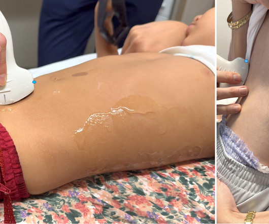
Take the ALiEMU PEM POCUS: Soft Tissue Quiz Case Goals List the indications of performing a pediatric soft tissue point-of-care ultrasound (POCUS). Pediatric Soft Tissue POCUS Ultrasound Technique Figure 1. Linear ultrasound transducer Probe Use a linear, high-frequency transducer. Describe the limitations of soft tissue POCUS.
Given her pain with a history of intermittent hematuria and dysuria, you perform a renal and bladder point of care ultrasound (POCUS) examination. Pre-warmed ultrasound gel is helpful when available. Then test your skills on the ALiEMU course page to receive your PEM POCUS badge worth 2 hours of ALiEMU course credit.
Background: Point-of-care ultrasound (PoCUS) is a valuable clinical tool in the assessment of acute dyspnea. Impact of serial cardiopulmonary point-of-care ultrasound exams in patients with acute dyspnoea: a randomized, controlled trial. PoCUS evaluations included lung ultrasound (LUS) and focused cardiac ultrasound (FoCUS).
Johnson, MD ( Community EM, Salina Regional Health Center) // Reviewed by: Joshua Lowe, MD (EM Attending Physician, USAF); Marina Boushra, MD (Cleveland Clinic Foundation, EM-CCM); Brit Long, MD (@long_brit) Case A 40-year-old woman presents to a rural emergency department (ED) with left leg pain and swelling for the past 5 days.
In these situations, the American Heart Association (AHA) and the European Resuscitation Council (ERC) of 2015 recommend the intraosseous (IO) route after the peripheral route and before the central venous route ( 1). 2015 PMID: 25768683 8 Leidel BA. This can often lead to significant delays in proper resuscitation. minutes CVC group.
Partial rupture of the proximal Achilles tendon: a differential diagnostic problem in ultrasound imaging. 2015; 3(4): PMID: 26665055 Post Peer Reviewed By: Salim R. Rezaie, MD (Twitter/X: @srrezaie ) The post REBEL Core Cast 116.0 – Achilles Tendon Rupture appeared first on REBEL EM - Emergency Medicine Blog. Sports Med.
The idea of the FOAMed review is to give you a digestible selection of reliable content from the online EM/CC world that you can fit into your busy weekly schedule. Over a year's span we will be sure to include topics from all core EM content areas.even the ones that may not be the coolest. A must listen here at EM Crit.
The idea of the FOAMed review is to give you a digestible selection of reliable content from the online EM/CC world that you can fit into your busy weekly schedule. Over a year's span we will be sure to include topics from all core EM content areas.even the ones that may not be the coolest. Find out here at Ultrasound Of The Week.
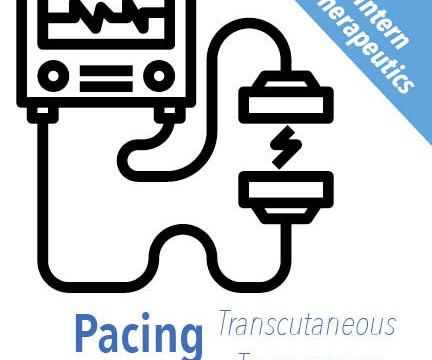
MedEdPORTAL Publications; 2015. Circulation , vol. 1103–1104, [link] Cousar J, Bohanske M, Hill J. Transvenous Cardiac Pacemaker Educational Resource. Available from: [link] Siamak Moayedi, Priya Patel, Nicholas Brady, Michael Witting, Timm-Michael L.
Background: The use of ultrasound is well established for trauma patients in the emergency department, with almost every patient receiving a FAST (Focused Assessment with Sonography in Trauma) examination as part of the “ABC’s” of trauma. Not so FAST- Chest ultrasound underdiagnoses traumatic pneumothorax. PMID: 34932040.
It might be better to consider traumatic cardiac arrest as a completely different disease eg LOST: Low Output State due to Trauma The 2015 European Resuscitation Council and UK Resuscitation Council Algorithms for Traumatic Cardiac Arrest: To read the whole ERC guideline on special circumstances cardiac arrest including trauma, click here.
The idea of the FOAMed review is to give you a digestible selection of reliable content from the online EM/CC world that you can fit into your busy weekly schedule. Over a year's span we will be sure to include topics from all core EM content areas.even the ones that may not be the coolest. Read the post from EM Nerd here.
Esophageal Balloon Tamponade Devices – Linton-Nachlas, Sengstaken-Blakemore, Minnesota Tubes (image courtesy of Dr. Mark Ramzy at REBEL EM) What are esophageal balloon tamponade devices? Additional trick: If the x-ray is delayed, you can pre-check with ultrasound [7]. Published July 13, 2015. Deranged Physiology.
The idea of the FOAMed review is to give you a digestible selection of reliable content from the online EM/CC world that you can fit into your busy weekly schedule. Over a year's span we will be sure to include topics from all core EM content areas.even the ones that may not be the coolest. Should we all be jumping on board?
Case A patient arrives via EMS from the bus station complaining of fever, vomiting, and back pain. Clinical impact: The patient’s DVT ultrasounds were negative. Our experience: It was not long ago that we instructed our staff that: ‘COWS >8, give ’em 8 (mg of buprenorphine).’ 2015 Apr 28;313(16):1636-44.
Guest Skeptic: Dr. Stephen Meigher is the EM Chief Resident training with the Jacobi and Montefiore Emergency Medicine Residency Training Program. Guest Skeptic: Dr. Stephen Meigher is the EM Chief Resident training with the Jacobi and Montefiore Emergency Medicine Residency Training Program. The TOMAHAWK Investigators.
EMS obtained the following vital signs: pulse 50, respiratory rate 16, blood pressure 96/49. It appears EMS obtained two EKGs, but unfortunately these were not saved in the medical record. The EMS crew was only BLS certified, so EKG interpretation is not within their scope of practice. Fortunately, that is exactly what happened.
Bagheri 2015 ). Signs Shafer’s sign (tobacco dust) May see a Weiss ring when the posterior vitreous (PV) detaches from the optic disc margin Visualization with ultrasound. 790-819 Core Ultrasound: Vitreous vs Retinal Detachmen t Post Created By: Anand Swaminathan MD, MPH Post Peer Reviewed By: Salim R.
For example, here are the locations identified as ‘2nd ICS mid clavicular line’ amongst 25 EM physicians in a 2005 EMJ paper. Ultrasound determination of chest wall thickness: implications for needle thoracostomy. Ultrasound in Emergency Medicine. Note – again please do not use this location! C., & MD, J.
It was edited by Smith CASE : A 52-year-old male with a past medical history of hypertension and COPD summoned EMS with complaints of chest pain, weakness and nausea. Smith comment: This patient did not have a bedside ultrasound. En route, EMS administered aspirin 325mg by mouth, but withheld nitroglycerin due to initial hypotension.
Jeff: And while it’s not exactly core EM, we’re going to briefly discuss indications for bariatric surgery, as this is something we don’t often review even in academic training programs. Which again reiterates why this is such an important topic for us as EM clinicians to be well-versed in. Jeff: Next up is ultrasound.
Record, 2015) Normal aortomesenteric angle ranges from 28 o to 65 o and the normal distance from 10 to 34 mm. Record, 2015) Nausea and pain are typically post-prandial. Record, 2015). Record, 2015) Secondary findings on imaging including gastric distension and distension of the proximal duodenum. The Oschner Journal.
link] PMID: 36553274 The post Congenital Pulmonary Airway Malformation (CPAM) appeared first on Pediatric EM Morsels. I know we already know this, but it especially true when you are talking about trying to obtain images on a wiggling fetus somewhere around the size of a banana. Dec: 9 (12): 1830.
We’ll keep it short, while you keep that EM brain sharp. A lower extremity radiograph does not reveal any gas formation and an ultrasound of the lower extremity is negative for DVT or cobblestoning, WBC is within normal limits and there are mild elevations in ESR and CRP. 2015, July 31). What is the most likely diagnosis?
Jeff: Edoxaban is up next, approved by the FDA in 2015 for similar indications as the other Factor Xa inhibitors. Interestingly, one retrospective study found limited agreement between EMS records and hospital documentation on current DOAC usage. But next we should talk about ultrasound.
The idea of the FOAMed review is to give you a digestible selection of reliable content from the online EM/CC world that you can fit into your busy weekly schedule. Over a year's span we will be sure to include topics from all core EM content areas.even the ones that may not be the coolest. Listen here @ EM Crit. Emlyns Blog.
Randomized, Controlled Trial of Ultrasound-Assisted Catheter-Directed Thrombolysis for Acute Intermediate-Risk Pulmonary Embolism. A prospective, Single-Arm Multicenter Trial of Ultrasound-Facilitated, Catheter-Directed, Low-Dose Fibrinolysis for Acute Massive and Submassive Pulmonary Embolism: The SEATTLE II Study. CHEST 2010.
Case: A 71-year-old man is brought to your emergency department (ED) by emergency medical serviced (EMS) having fallen two steps at home. EMS have already splinted an obvious mid-shaft femoral fracture, but he continues to be tachycardic and hypotensive.
Author: Kristine Jeffers, MD ( EM Physician , San Antonio Uniformed Services Health Education Consortium) // Reviewed by: Jessica Pelletier, DO (EM Education Fellow, Washington University in St. Half of all embolisms occur within the first 24 hours and 75% will present by postoperative day 3. 2020 Sep;127(10):1269-1279.
If you want to know about ultrasound findings of pneumothorax, check out this great R.E.B.E.L. 2015; 261: 1068-78. And you can see what a CT scan of a patient with tension pneumothorax looks like in this vimeo shared on the Life in the Fast Lane website. References: Leigh-Smith S, Harris T. Tension pneumothorax—time for a re-think?
My bedside ultrasound was of insufficient quality, but showed somewhat reduced overall EF, distended IVC without respiratory variation, no pericardial effusion, and diffuse bilateral B lines. == What do you think of her ECG?
Advocacy for Trauma Care and EMS Development. Development of EM Residencies. Advances in Medical Imaging (CT, MRI, and Ultrasound) facilitating rapid, accurate, diagnosis affording new potential to save additional lives. Organized EM supporting Rapid Sequence Intubation and other airway advances. EDNA->ENA.
Thankfully, fatalities are declining, with just 565 in 2015. Jeff: With your initial stabilization underway, you can begin to gather a more thorough history either from bystanders or EMS if they are still present. Jeff: Real quick – in case you missed it – ultrasound sneaks in again. Let’s move on to treatment.
CT is good but you really should learn ultrasound, and lastly, sick patients need prompt consultation and resuscitation, not rapid trips to radiology. Nachi: Diagnosis of appendicitis, in a pregnant patient, ultrasound vs. mri. Jeff: Next we have everybody’s favorite, the ultrasound. Jeff: Indeed. Sounds familiar.
Nachi: And if the prehospital team is lucky enough, or maybe unlucky enough, i don’t know, to have a credentialed provider who can perform ultrasound for those suspected of having a blunt cardiac injury, the general prehospital data on ultrasound is sparse. Jeff: Great, let’s move onto ED care, beginning with the H&P.
Nachi: Sometimes… Jeff: This month’s issue was authored by Mollie Williams, who is the EM residency program director at the Brooklyn Hospital Center. Acute effects of synthetic cannabinoids: update 2015. Cannabinoids for medical use: a systematic review and meta-analysis. 2015;313(24):2456-2473. Retrospective chart review; 4 cases) 64.
As some background, from 2006-2015 there were almost 66,000 reported snake exposures and 31 deaths from snake envenomation in the US. If EMS has placed bandages, leave them in place until antivenom and resuscitative equipment is ready. Jeff: What a team! But, let’s get back to the snakes. Jeff: What a team!
I know this is a HOTLY debated topic among EM Docs. So to get us all on the same page, the discriminatory zone is the b-HCG at which an IUP is expected to be seen on ultrasound. Nachi: Definitely concerning, but this is all the more reason you need to employ our favorite imaging modality… the ultrasound.
Check : [vitals, SOB, Chest Pain, Ultrasound] If the patient has Abdominal Pain, Chest Pain, Dyspnea or Hypoxemia, Headache, Hypotension , then these should be considered the primary chief complaint (not syncope). Aortic Dissection, Valvular (especially Aortic Stenosis), Tamponade. Good History and Physical exam, including a. Quinn et al.
We organize all of the trending information in your field so you don't have to. Join 5,000+ users and stay up to date on the latest articles your peers are reading.
You know about us, now we want to get to know you!
Let's personalize your content
Let's get even more personalized
We recognize your account from another site in our network, please click 'Send Email' below to continue with verifying your account and setting a password.


Let's personalize your content