This site uses cookies to improve your experience. To help us insure we adhere to various privacy regulations, please select your country/region of residence. If you do not select a country, we will assume you are from the United States. Select your Cookie Settings or view our Privacy Policy and Terms of Use.
Cookie Settings
Cookies and similar technologies are used on this website for proper function of the website, for tracking performance analytics and for marketing purposes. We and some of our third-party providers may use cookie data for various purposes. Please review the cookie settings below and choose your preference.
Used for the proper function of the website
Used for monitoring website traffic and interactions
Cookie Settings
Cookies and similar technologies are used on this website for proper function of the website, for tracking performance analytics and for marketing purposes. We and some of our third-party providers may use cookie data for various purposes. Please review the cookie settings below and choose your preference.
Strictly Necessary: Used for the proper function of the website
Performance/Analytics: Used for monitoring website traffic and interactions
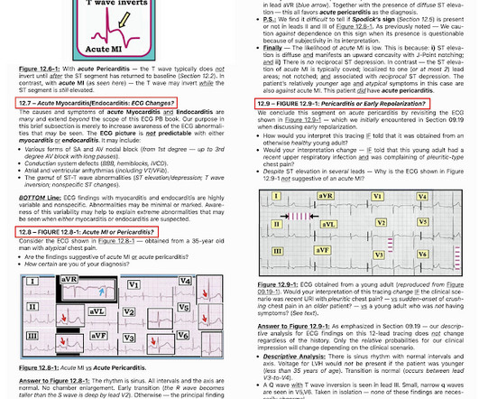
The ECG in Figure-1 — was obtained from a middle-aged woman with positional tachycardia and diaphoresis with change of position from suprine to sitting. Although CP ( C hest P ain ) was not a prominent symptom — ACS ( A cute C oronary S yndrome ) was suspected from the chest lead T wave inversion seen on this ECG. WHY — or Why Not?
The ECG in Figure-1 was obtained from a man in his 40s — who presented to the ED ( E mergency D epartment ) because of CP ( C hest P ain ) and shortness of breath. QUESTIONS: In view of the above history — How would YOU interpret the ECG in Figure-1 ? Based on the history and the patient's initial ECG — the cath lab was activated.
For full discussion of this case — See ECG Blog #191 — == The 2-lead rhythm strip shown in Figure-1 was obtained from an elderly woman who presented to the ED following a syncopal episode. ECG Media Pearl # 8 ( 8:20 minutes Video ) — ECG Blog #191 — Distinguishing between A V D issociation vs Complete AV Block ( 2/6/2021 ).
Figure-1: The initial ECG in today's case. KEY Clinical Point: If I was the medical provider charged with the care of the patient whose ECG is shown in Figure-1 — I would approach this tracing in the following sequential stages: I’d first establish that the patient was hemodnamically stable with this ECG and this cardiac rhythm.
The ECG in Figure-1 was obtained from a woman in her 60s — who was seen in the ED ( E mergency D epartment ) as part of her evaluation for trauma following a motor vehicle accident. Figure-1: The initial ECG in today's case. To do this — I apply the P s, Q s, 3 R Approach ( See ECG Blog #185 — for review of my system ).
I was sent the ECG in Figure-1 — told only that the patient was 70 years old, and had a history of an ASD ( A trial S eptal D efect ). The patient was hemodynamically stable with ECG #1. Figure-1: The initial ECG in today's case. Serum K+ was normal. QUESTIONS: How would YOU interpret the rhythm in Figure-1 ? Are there P waves?
The 12-lead ECG and long lead II rhythms shown in Figure-1 — was obtained from an older man with a recent history of “easy fatiguability” and a presyncopal episode. QUESTIONS: How would YOU interpret the ECG in Figure-1 ? Figure-1: The initial ECG in today’s case. I outline my approach for doing so below.
Imagine the only information provided for the ECG in Figure-1 — is that it was obtained from a 60-year old man with new CP ( C hest P ain ). QUESTIONS: In view of this brief history — How would YOU interpret this ECG in Figure-1 ? Is the cardiac rhythm related to the 12-lead ECG? Figure-1: The initial ECG in today’s case.
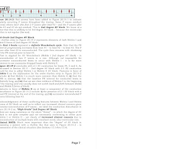
For full discussion of this case — See ECG Blog #344 — == How would YOU interpret the lead II rhythm strip shown in Figure-1 ? Section 2F ( 6 pages = the " short " Answer ) from my ECG-2014 Pocket Brain book provides quick written review of the AV Blocks ( This is a free download ). Or — Is it “ something else ”?
I was sent the tracing shown in Figure-1 — told only that this was a preoperative ECG obtained from an asymptomatic older woman scheduled for non-cardiac surgery. How would YOU interpret this ECG? Figure-1: Preoperative ECG from an asymptomatic older woman scheduled for non-cardiac surgery. The 12-lead ECG is very concerning.
You are asked to interpret the ECG in Figure-1. What is the rhythm in ECG #1 ? MY Thoughts on the ECG in Figure-1: I routinely begin assessment of each 12-lead ECG I encounter — with interpretation of the rhythm. To do this — I apply the P s, Q s, 3 R Approach ( See ECG Blog #185 — for review of my system ).
The ECG in Figure-1 is from a man in his 30s — who overall has been healthy, except for a history of "intermittent palpitations" that he has had since childhood. He was hemodynamically stable with ECG #1. Figure-1: The initial ECG in today's case. To improve visualization — I've digitized the original ECG using PMcardio ).
The ECG in Figure-1 was obtained from an elderly woman — who presented to the ED ( E mergency D epartment ) for dyspnea on exertion over recent weeks. Figure-1: The initial ECG in today's case. ( To improve visualization — I've digitized the original ECG using PMcardio ). What are YOUR "Quick Thoughts" about this case?
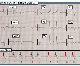
The ECG in Figure-1 was obtained from a man in his mid-60s — who presented with new chest pain. Figure-1: T he initial ECG in today’s case. MY Thoughts on the Initial ECG: The rhythm in ECG #1 — is sinus at ~70/minute. Figure-2: I've labeled significant ST-T wave findings in the limb leads of ECG #1.
You are given this ECG to review. QUESTION: Is there a potential problem with this ECG? Figure-1: The initial ECG in today's case. == N OTE : Many of us are charged with reviewing ECGs that have been interpreted by other clinicians — often without the benefit of much ( or any ) history. The rhythm in ECG #1 is sinus.
The ECG in Figure-1 was obtained from a previously healthy 30-ish year old man — who presented to medical attention for vasovagal syncope. Based on this initial ECG — the patient was transferred to a PCI-capable center: Do YOU agree with the need for transfer? Figure-1: The initial ECG in today's case.
The 12-lead ECG and long lead rhythm strip in Figure-1 — was obtained from a previously healthy 15-year old male , who presented with fever and diarrhea. How would YOU interpret the ECG in Figure-1 ? Figure-1: The initial ECG in today’s case — obtained from a 15-year old male with fever and diarrhea. No chest pain.
No EKG arrived with the transfer packet, so I ordered a STAT EKG and went bedside to see her. I walked into the room to see the following EKG before I had the chance to ask any questions. EKG 1 What do you think? We would have taught her "acute reperfused" for this ECG. The angiography fits perfectly with the EKG.
Written by Pendell Meyers Try first to interpret the ECG without any clinical context: What do you think? Overall, this looks like one of the rare ECGs that is actually specific for pericarditis in my opinion. There was no prior ECG for comparison. To improve visualization — I've digitized the original ECG using PMcardio ).
What do you think of the ECG, and does it matter? I sent this to the Queen of Hearts So the ECG is both STEMI negative and has no subtle diagnostic signs of occlusion. 2] This is because, contrary to Bayesian reasoning, the STEMI paradigm is named after and defined by one part of one test: ST elevation on ECG. But only 6.4%
Her ECG is shown below: What do you think? The conventional machine algorithm interpreted this ECG as STEMI. Alternatively, with STE in V1 and III, and STD in I and aVL, this ECG could represent proximal RCA OMI with right ventricular involvement. What do you do clinically when the ECG looks like this?
Check the pulse RSI= Resuscitation Sequence Intubation Hypoxia, Hypotension, and Acidosis are the reason patients code during/post intubation These patients are super high risk for all 4 Optimize first pass success – Induction agent + paralytic Unconscious patients will still have muscle tone Induction Ketamine or Etomidate at half doses (i.e.,
This week’s ECG is from a 62 yr old male who presented complaining of palpitations for the preceding 4 hours. See the ECG at: ECG of the Week &#… No significant medical history or medications.
This week’s ECG case is a rhythm strip from a 77yr old male. He complained of chest pain during this ECG recording. He has a history of hypertension and hypercholesterolaemia. Check out the E…
The following ECG was obtained. Note that the machine read is "normal sinus rhythm, normal ECG." ECG 1 What do you think? I sent this ECG to Dr. Smith and Dr. Meyers with no clinical context. Smith comment: this troponin alone should be enough data to activate the cath lab, regardless of the ECG. <0.049 ng/mL).
Clinical conclusion and interpretation on last week’s ECG case of a middle aged male who presented to the Emergency Department with … Continue reading →
This week’s case had a 97yr old presenting with presyncope, and 2 ECG’s performed only 1 minute apart. Check out the interpretation … Continue reading →
A 51yr old presents with a post-prandial episode of syncope and subsequent sensation of episodic pre-syncope. No relevant past medical … Continue reading →
Are you looking for a comprehensive ECG glossary that goes beyond simply defining words? Dr. Ken Grauer, who is the ECG Guru's Consulting Expert, has a Glossary available on his website that explains the terms. The glossary is exerpted from his e-Publication, "A 1st Book On ECGs - 2014", available on Amazon.
Remove ECG leads and patches. Reattach EKG leads to back. Place a new bed sheet on the side of the bed that the patient will face when in this lateral decubitus position. Leave most of the sheet hanging. Turn patient into lateral decubitus position. Suction as needed. Log-roll the patient into the prone position. 2013; Mi et.
I want all to know that, with the right mind preparation, and the use of the early repol/LAD occlusion formula, extremely subtle coronary occlusion can be detected prospectively, with no other information than the ECG. His ECG was repeated at this point: This shows a well developed anterior STEMI. His first troponin was normal.
This year we are very fortunate to have Dr. Amal Mattu, EKG Jedi, as teaching faculty for our 39th Maine Medical Center/Maine ACEP Winter Symposium. HR 137, BP 105/50, 36C, RR 26, 95% O2 saturation on room air As he is brought in by ambulance, the paramedic presents the prehospital ECG below… do you activate the cath lab? Pubmed ]
Here was his ED ECG: I read this as normal --One might say there is ST depression in II, III, and aVF, but this is merely an atrial repolarization wave. --You We recorded a posterior ECG: V4-V6 are moved around to the back and are really V7-V9. Learning Point Acute coronary occlusion may occur with no ECG findings whatsoever.
This ECG was presented in a conference. I was asked to interpret his ECG in the conference. ECG-1: What do you think I said? He had no more ECGs recorded. Here is data from a study we published in 2014 for type II NonSTEMI: Sandoval Y. ECG-2 Now it is more obvious. I had never seen it before. First was 2.9
Here is his ECG ( Figure 1 ): What do you think? There may be ischemia present, but it is not evident on the ECG. Importantly, all ST-T abnormalities are discordant to (in the opposite direction of) the majority of the QRS) Indeed, this was the patient's baseline ECG. Journal of Electrocardiology 47 (2014) 655–660.
We organize all of the trending information in your field so you don't have to. Join 5,000+ users and stay up to date on the latest articles your peers are reading.
You know about us, now we want to get to know you!
Let's personalize your content
Let's get even more personalized
We recognize your account from another site in our network, please click 'Send Email' below to continue with verifying your account and setting a password.


Let's personalize your content