EM@3AM: Brainstem Strokes
EMDocs
MAY 11, 2024
Answer : Brainstem stroke specifically in the pons resulting in locked in syndrome. CT head without contrast 1 is performed and reveals the following: Question: What is the diagnosis?

EMDocs
MAY 11, 2024
Answer : Brainstem stroke specifically in the pons resulting in locked in syndrome. CT head without contrast 1 is performed and reveals the following: Question: What is the diagnosis?

RebelEM
FEBRUARY 24, 2025
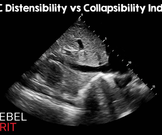
Of course, there are other methods of assessing fluid tolerance : Capillary refill evaluation, passive leg raise, central venous pressure measurement, pulmonary artery wedge pressures, stroke volume variation, pulse pressure variation, etc. Ultrasound Med Biol. Most measurements are somewhere around the hepatic confluence.
This site is protected by reCAPTCHA and the Google Privacy Policy and Terms of Service apply.

RebelEM
JANUARY 29, 2024
Randomized, Controlled Trial of Ultrasound-Assisted Catheter-Directed Thrombolysis for Acute Intermediate-Risk Pulmonary Embolism. A prospective, Single-Arm Multicenter Trial of Ultrasound-Facilitated, Catheter-Directed, Low-Dose Fibrinolysis for Acute Massive and Submassive Pulmonary Embolism: The SEATTLE II Study. Am J Cardiol 2013.

Taming the SRU
FEBRUARY 5, 2024
It is most helpful to do the ultrasound immediately before needle insertion, as movement of the patient may shift cutaneous landmarks from underlying bony structures. This resource offers additional information on ultrasound assisted LP’s. WHY - Why Not? REFERENCES 1. Zambito Marsala, S., Gioulis, M., & Pistacchi, M. Glimåker, M.,

RebelEM
MARCH 8, 2023
CRAO is a stroke of the eye; patients should be considered for a complete stroke work up. Signs Shafer’s sign (tobacco dust) May see a Weiss ring when the posterior vitreous (PV) detaches from the optic disc margin Visualization with ultrasound. Philadelphia: Elsevier, 2013 (Ch) 26: p. >10 floaters, (+) LR 8.1-36

Pediatric Emergency Playbook
OCTOBER 1, 2017
Also used for severe MS, stroke, TBI, chronic pain. 2013; 49:138–144. Ultrasound-guided refilling of an intrathecal baclofen pump—a case report. 2013; 29:347–349. Are VNS safe in everyday life? Verify the medication and identify the toxidrome if symptomatic. Pediatr Neurosurg. Yang TF, Wang JC, Chiu JW et al.

PulmCCM
JULY 10, 2023
Read in JAMA When to start anticoagulation after ischemic stroke from atrial fibrillation It’s been unclear when to start anticoagulation after an ischemic stroke presumed due to thromboembolism from atrial fibrillation. Aspirin is sometimes given during the waiting period. Heparin infusions are usually avoided.
Let's personalize your content