Remove 2013 Remove Emergency Department Remove Ultrasounds
article thumbnail

Ultrasound of the Month: Peritonsillar Abscess

Taming the SRU

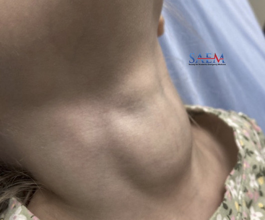
THE CASE A female in her 20’s presented to the emergency department with concern for oral swelling. IMAGING WITH ULTRASOUND Peritonsillar abscess is one of the most common deep space infections of the head and neck contributing significantly to health care costs in the United States. There was no trismus.

article thumbnail

Predictive values of indirect ultrasound signs for low risk of acute appendicitis in paediatric patients without visualisation of the appendix on ultrasound

Emergency Medicine Journal

Background and objectives The ability to rule appendicitis in or out using ultrasound is limited by studies where the appendix is not visualised. For those patients who had formal ultrasound, direct and indirect findings of ultrasound were abstracted from the ultrasound report. 95% CI 1.7 95% CI 3.8 95% CI 1.4

Insiders

Sign Up for our Newsletter

This site is protected by reCAPTCHA and the Google Privacy Policy and Terms of Service apply.

Trending Sources

article thumbnail

Diagnostics and Therapeutics: Vascular Access in the Emergency Department

Taming the SRU

Ultrasound Guided Peripheral IV (USGIV) Access anatomy of arm veins when considering USGIV. unported license [link] Ultrasound allows us to visualize deeper, typically larger veins in the forearm and upper arm. Place ultrasound machine on the opposite side of the bed to minimize neck strain. Check for flash in the IV chamber.

article thumbnail

SGEM#415: Buckle Down for some Ultrasound to Diagnosis Distal Forearm Fractures

The Skeptics' Guide to EM

Casey Parker is a Rural Generalist that includes in his practice emergency medicine, anesthesia, and critical care. Casey currently splits his time […] The post SGEM#415: Buckle Down for some Ultrasound to Diagnosis Distal Forearm Fractures first appeared on The Skeptics Guide to Emergency Medicine. Reference: Snelling et al.

Fractures 124
article thumbnail

PEM POCUS Series: Pediatric Renal and Bladder Ultrasound

ALiEM

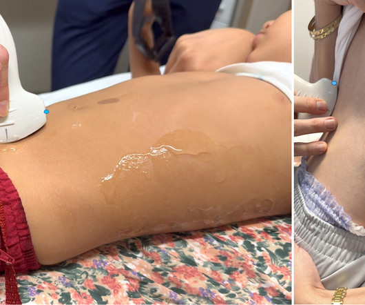
Given her pain with a history of intermittent hematuria and dysuria, you perform a renal and bladder point of care ultrasound (POCUS) examination. Pre-warmed ultrasound gel is helpful when available. Then test your skills on the ALiEMU course page to receive your PEM POCUS badge worth 2 hours of ALiEMU course credit.

article thumbnail

Mastering Minor Care: Knee Arthrocentesis

Taming the SRU

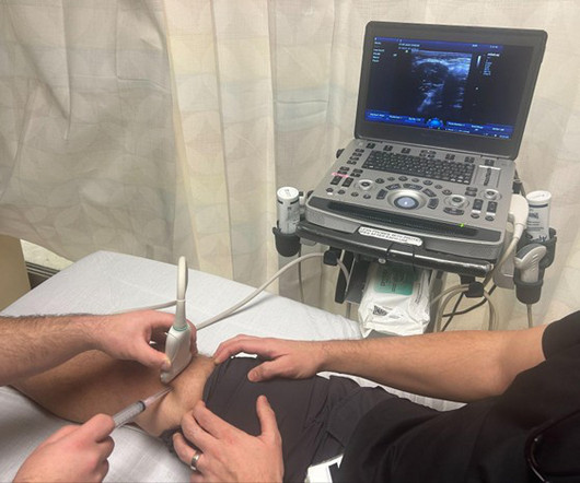
INDICATIONS Arthrocentesis is a common procedure performed in the Emergency Department, in which a needle is inserted into a joint to withdraw fluid from the joint capsule. Studies have shown that effusions of as little as 2 to 4 mL can be identified using ultrasound [6]. ANESTHETIZE THE AREA step 7. Step 1: Prepare STEP 2.

article thumbnail

Serial PoCUS for ED Patients with Acute Dyspnea: Is More Actually Better?

RebelEM

Background: Point-of-care ultrasound (PoCUS) is a valuable clinical tool in the assessment of acute dyspnea. In many emergency departments (ED), US machines are readily available and can be used to rapidly assess and monitor patients with acute dyspnea at the bedside. Emerg Med J. 2023;40(10):700-707.