Remove 2013 Remove EKG/ECG Remove Wellness
article thumbnail

ECG Blog #402 — Will Adenosine Convert This?

Ken Grauer, MD

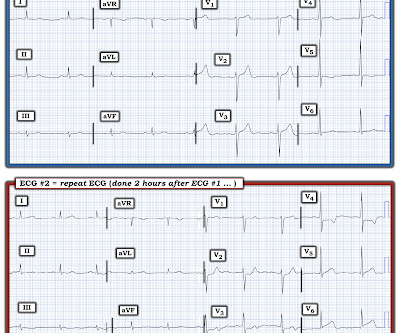
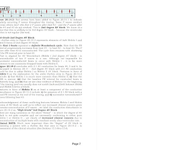
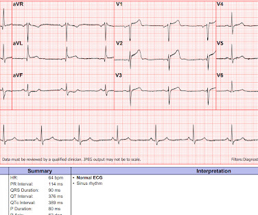
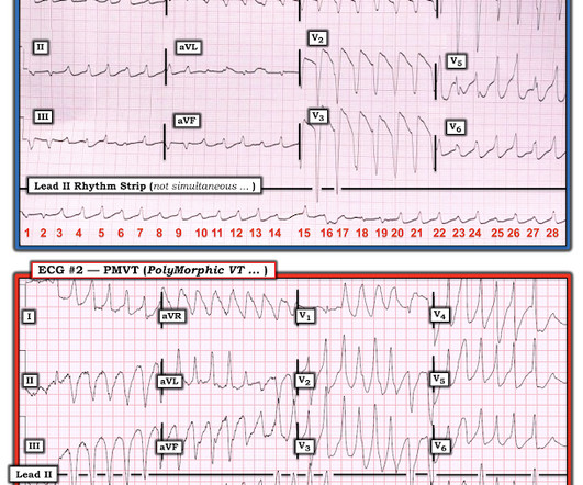
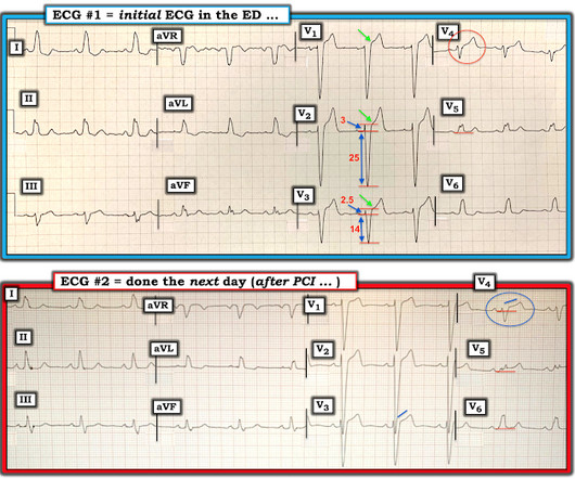
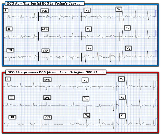
Figure-1: How would YOU interpret this ECG? MY Thoughts on the ECG in Figure-1: When faced with a challenging cardiac arrhythmia — It is a "luxury" to have access to a long lead rhythm strip containing 3 simultaneously -recorded leads. The repeat ECG after this treatment is shown in Figure-4.

EKG/ECG 419
article thumbnail

ECG Blog #411 — Is it Wenckebach?

Ken Grauer, MD

Figure-1: The initial ECG in today's case. KEY Clinical Point: If I was the medical provider charged with the care of the patient whose ECG is shown in Figure-1 — I would approach this tracing in the following sequential stages: I’d first establish that the patient was hemodnamically stable with this ECG and this cardiac rhythm.

EKG/ECG 441
Insiders

Sign Up for our Newsletter

This site is protected by reCAPTCHA and the Google Privacy Policy and Terms of Service apply.

Trending Sources

article thumbnail

ECG Blog #415 — The Cath showed NO Occlusion!

Ken Grauer, MD

The ECG in Figure-1 was obtained following successful resuscitation. QUESTIONS: In view of the above history — How would YOU interpret the ECG in Figure-1 ? Is this ECG finding present in today’s initial ECG? Figure-1: The initial ECG in today's case — obtained after successful resuscitation from cardiac arrest. (

EKG/ECG 408
article thumbnail

ECG Blog #416 — Is the Rhythm and ECG related?

Ken Grauer, MD

Imagine the only information provided for the ECG in Figure-1 — is that it was obtained from a 60-year old man with new CP ( C hest P ain ). QUESTIONS: In view of this brief history — How would YOU interpret this ECG in Figure-1 ? Is the cardiac rhythm related to the 12-lead ECG? Figure-1: The initial ECG in today’s case.

EKG/ECG 262
article thumbnail

ECG Blog #401 — What Kind of Block?

Ken Grauer, MD

The ECG in Figure-1 was obtained from an elderly woman — who presented to the ED ( E mergency D epartment ) for dyspnea on exertion over recent weeks. Figure-1: The initial ECG in today's case. ( To improve visualization — I've digitized the original ECG using PMcardio ). What are YOUR "Quick Thoughts" about this case?

EKG/ECG 254
article thumbnail

Concerning EKG with a Non-obstructive angiogram. What happened?

Dr. Smith's ECG Blog

It appears EMS obtained two EKGs, but unfortunately these were not saved in the medical record. The EMS crew was only BLS certified, so EKG interpretation is not within their scope of practice. The patient arrived just after 10 AM, and the following EKG was obtained. There are no further EKGs or troponin measurements.

EKG/ECG 127
article thumbnail

ECG Blog #377 — Is the 1st ECG Normal?

Ken Grauer, MD

The patient whose initial ECG is shown in Figure-1 — is a middle-aged man who presented to the ED ( E mergency D epartment ) for new-onset CP ( C hest P ain ). QUESTIONS: In view of the above history — How would YOU interpret the initial ECG? WHY is it important to correlate severity of this patient’s CP with ECG #1 ?

EKG/ECG 195