Remove 2013 Remove EKG/ECG Remove Shock
article thumbnail

ACMT Toxicology Visual Pearl: Salt, not Shock

ALiEM

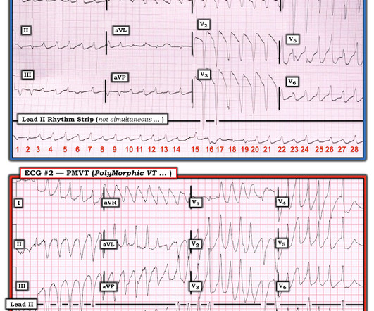
What agent would most likely be responsible for these ECG findings? This EKG shows a wide complex (QRS 240 msec), irregular rhythm with left bundle branch block morphology at a rate slower than expected (90 bpm) for a ventricular arrhythmia such as ventricular tachycardia. 2013 Dec;15(4):90-2. Epub 2013 Nov 7.

Shock 73
article thumbnail

Chemical Burns

Mind The Bleep

Circulation Assess heart rate, blood pressure, peripheral and central CRT, pulses and 3 lead ECG. Sodium Chloride or Hartmanns if indicated, monitoring for signs of shock. 2013 May;74(5):1363-6. Establish IV access and begin fluid resuscitation with 250ml boluses of 0.9% Advanced trauma life support (ATLSĀ®): the ninth edition.

Burns 52
Insiders

Sign Up for our Newsletter

This site is protected by reCAPTCHA and the Google Privacy Policy and Terms of Service apply.

Trending Sources

article thumbnail

Diabetic Ketoacidosis in Paediatrics

Mind The Bleep

ECG: to monitor T wave changes due to hypokalaemia. ECG features of Hypokalaemia: Increased P wave amplitude (peaked P waves) Prolonged PR interval Widespread ST depression T wave flattening or inversion Prominent U waves (most noticeable in the precordial leads) Figure 2 : ECG of a patient with serum K+ of 1.9

article thumbnail

Diffuse Subendocardial Ischemia on the ECG. Left main? 3-vessel disease? No!

Dr. Smith's ECG Blog

An immediate 12-lead EKG was obtained: There is ST elevation in leads aVR and V1, with marked ST depression in I, II, III, aVF, V3-V6. DISCUSSION: The 12-lead EKG EMS initially obtained for this patient showed severe ischemia, with profound "infero-lateral" ST depression and reciprocal ST elevation in lead aVR. What should be done?

EKG/ECG 40
article thumbnail

Episode 7: Sepsis

PHEM Cast

Think about ECG as an example, So, where the box is green, the test has given us the correct result for the patient. One year mortality of patients treated with an emergency department based early goal directed therapy protocol for severe sepsis and septic shock: a before and after study. 2013 Jun;44(6):1116ā€“25. J Emerg Med.

Sepsis 52
article thumbnail

90 year old with acute chest and epigastric pain, and diffuse ST depression with reciprocal STE in aVR: activate the cath lab?

Dr. Smith's ECG Blog

What is your ECG interpretation and what would you do next? This ECG shows a normal sinus rhythm with a normal conduction pattern (normal PR, normal QRS, and normal QTc), normal axis, late R wave progression (and misplaced V2), normal voltages, ST-elevation in aVR and global ST-depressions. BP was 110 and oxygen saturation was normal.

EKG/ECG 52
article thumbnail

A Different Kind of Wide Rhythm -- Pleomorphic Ventricular Tachycardia

Dr. Smith's ECG Blog

== MY Comment by K EN G RAUER, MD ( 6/1 /2020 ): == YOU are asked to interpret the ECG shown in Figure-1. Figure-1: The initial ECG in the ED. MY THOUGHTS on ECG #1: My initial impression on looking at the ECG in Figure-1 ā€” was that the rhythm was either rapid AFib in a patient with WPW ā€” or ā€” PMVT ( P oly M orphic VT ).

EKG/ECG 52