Remove 2013 Remove EKG/ECG Remove Outcomes
article thumbnail

A man in his early 40s with chest pain a "normal ECG" by computer algorithm. Should we avoid interrupting a physician to interpret his ECG?

Dr. Smith's ECG Blog

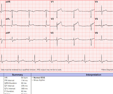
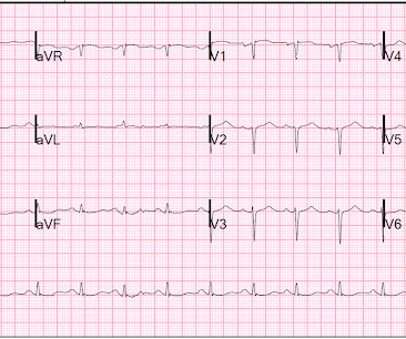
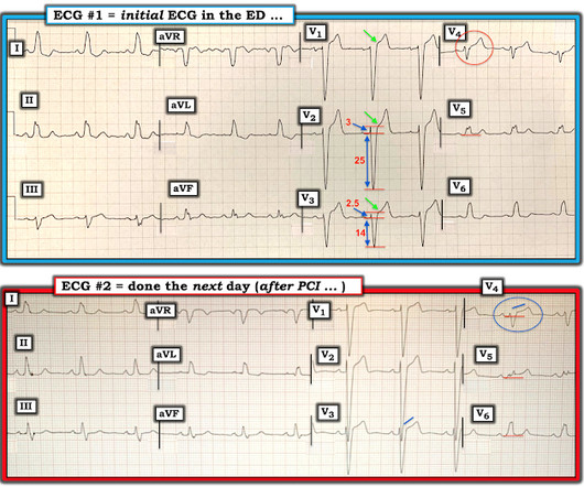
EMS arrived and recorded this ECG: What do you think? See same ECG below with computer automated interpretation, using the Glasgow ECG algorithm which apparently is used by many different providers and devices Amazing that the computer calls this normal. Next day ECG. And yet it still says "normal". Learning Points: 1.

EKG/ECG 98
article thumbnail

Where did the P waves go?

Dr. Smith's ECG Blog

Written by Willy Frick I was reading through ECGs in the queue when I saw the following. At first glance there seems to be a lot of variation happening, but by focusing on one part of the ECG at a time we can make sense of it. So why does the sinus node apparently stop pacing for a few seconds in the middle of the ECG?

EKG/ECG 110
Insiders

Sign Up for our Newsletter

This site is protected by reCAPTCHA and the Google Privacy Policy and Terms of Service apply.

Trending Sources

article thumbnail

A Tough ECG, But Learn From It!

Dr. Smith's ECG Blog

I want all to know that, with the right mind preparation, and the use of the early repol/LAD occlusion formula, extremely subtle coronary occlusion can be detected prospectively, with no other information than the ECG. His ECG was repeated at this point: This shows a well developed anterior STEMI. His first troponin was normal.

EKG/ECG 52
article thumbnail

Chemical Burns

Mind The Bleep

Circulation Assess heart rate, blood pressure, peripheral and central CRT, pulses and 3 lead ECG. Proper stabilization and detailed communication with the burns centre are essential for optimal patient outcomes. 2013 May;74(5):1363-6. Administer high-flow oxygen if indicated and consider arterial blood gas ( ABG ).

Burns 52
article thumbnail

Chest pain with 2 serial ECGs, with dynamic change, texted to me

Dr. Smith's ECG Blog

Queen: #1: NOT OMI, HIGH CONFIDENCE Queen: #2: NOT OMI, HIGH CONFIDENCE ECG 1 Interpretation: there is terminal T-wave in V3-V6. See 2 dozen examples here: Understanding this pathognomonic ECG would have greatly benefitted the patient. Outcome: Patient ruled out for MI by troponins. Is this Wellens' pattern A?

EKG/ECG 52
article thumbnail

What does the angiogram show? The Echo? The CT coronary angiogram? How do you explain this?

Dr. Smith's ECG Blog

She has no previous cardiac history of which she is aware 911 was called and here is her Prehospital ECG: What do you think? link] Case continued She arrived in the ED and here is the first ED ECG. In comparison with the first ECG, I would guess that the artery is reperfusing. Figure-1: The initial ECG in today's case.

EKG/ECG 109
article thumbnail

EM@3AM: Brainstem Strokes

EMDocs

Semin Ultrasound CT MR. 2013 Apr;34(2):131-41. Endovascular and Clinical Outcomes of Vertebrobasilar Intracranial Atherosclerosis-Related Large Vessel Occlusion. Reference article, Radiopaedia.org (Accessed on 28 Aug 2023) [link] Ortiz de Mendivil A, Alcalá-Galiano A, et al. doi: 10.1053/j.sult.2013.01.004. 2013.01.004. PMID: 23522778.]

Stroke 111