Remove 2013 Remove EKG/ECG Remove Hyperthermia / Hypothermia
article thumbnail

Chemical Burns

Mind The Bleep

Circulation Assess heart rate, blood pressure, peripheral and central CRT, pulses and 3 lead ECG. Exposure Expose the patient in a systematic manner while keeping remaining body areas covered e.g. 1 limb at a time, to reduce the risk of hypothermia. 2013 May;74(5):1363-6. Assess pupillary reaction to light. doi: 10.1097/TA.0b013e31828b82f5.

Burns 52
article thumbnail

Diabetic Ketoacidosis in Paediatrics

Mind The Bleep

ECG: to monitor T wave changes due to hypokalaemia. ECG features of Hypokalaemia: Increased P wave amplitude (peaked P waves) Prolonged PR interval Widespread ST depression T wave flattening or inversion Prominent U waves (most noticeable in the precordial leads) Figure 2 : ECG of a patient with serum K+ of 1.9 Marcdante, R.

Insiders

Sign Up for our Newsletter

This site is protected by reCAPTCHA and the Google Privacy Policy and Terms of Service apply.

Trending Sources

article thumbnail

emDOCs Revamp: Alcohol Withdrawal

EMDocs

2013; 88(9): 589-595. fold higher risk of NSTI than the control group 12 For those without comorbidities , AUD exhibited a 15.2-fold fold higher risk of NSTI than the control group 12 For those without comorbidities , AUD exhibited a 15.2-fold Alcohol withdrawal syndrome in medical patients. Cleve Clin J Med. 2016; 83(1): 67-79.

Seizures 105
article thumbnail

Hyperthermia and ST Elevation

Dr. Smith's ECG Blog

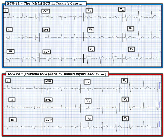
An initial EKG was obtained: Computer read: sinus tachycardia, early acute anterior infarct. Here is her prior EKG: When compared to the old EKG – Q waves present before, TWI in aVR present before, but all other changes are new. What is the differential for this EKG? T wave inversion III, aVR, TWF in aVF. Is this an OMI?

article thumbnail

A man in his 70s with weakness and syncope

Dr. Smith's ECG Blog

An ECG was performed in the ED at 1554: Original image unavailable, this is the only recorded scanned ECG available. In a patient with syncope and fever, this ECG looks more like Brugada. Smith comment: the ECG in question could be due to Brugada, even though there is a change from baseline. PM Cardio digitized version.

EKG/ECG 91
article thumbnail

A Middle-Aged male with Chest Pain and an Unusual ECG

Dr. Smith's ECG Blog

This ECG was sent from South Asia. Here is the ECG: What do you think? Followup ECG: No Change Absence of evolution is the best evidence against ischemia as the etiology. Could this be a coincidence that the patient was experiencing chest pain and upon ECG capture the atypical brugada pattern was found? Did it subside?

EKG/ECG 52
article thumbnail

Another deadly and confusing ECG. Are you still one of the many people who will be fooled by this ECG, or do you recognize it instantly?

Dr. Smith's ECG Blog

Here is the initial ED ECG: What do you think? The ECG shows peaked T waves, QRS widening, a coved-type ST segment with inverted T waves most prominent in V1 and V2 consistent with Brugada pattern. They recommended repeating his ECG and awaiting troponin since the patient did not have any chest pain. Creatinine elevated at 3.09

EKG/ECG 52