This site uses cookies to improve your experience. To help us insure we adhere to various privacy regulations, please select your country/region of residence. If you do not select a country, we will assume you are from the United States. Select your Cookie Settings or view our Privacy Policy and Terms of Use.
Cookie Settings
Cookies and similar technologies are used on this website for proper function of the website, for tracking performance analytics and for marketing purposes. We and some of our third-party providers may use cookie data for various purposes. Please review the cookie settings below and choose your preference.
Used for the proper function of the website
Used for monitoring website traffic and interactions
Cookie Settings
Cookies and similar technologies are used on this website for proper function of the website, for tracking performance analytics and for marketing purposes. We and some of our third-party providers may use cookie data for various purposes. Please review the cookie settings below and choose your preference.
Strictly Necessary: Used for the proper function of the website
Performance/Analytics: Used for monitoring website traffic and interactions
EMS obtained the following vital signs: pulse 50, respiratory rate 16, blood pressure 96/49. It appears EMS obtained two EKGs, but unfortunately these were not saved in the medical record. The EMS crew was only BLS certified, so EKG interpretation is not within their scope of practice.
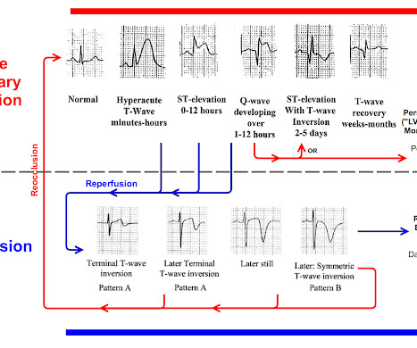
The ECG in Figure-1 was obtained from an elderly man with a history of coronary disease — who contacted EMS for "burning" chest discomfort that woke him at 3am. QUESTIONS: How would YOU interpret the initial ECG in today's case? In view of the above history — Does ECG #1 suggest an acute event?
We’ll keep it short, while you keep that EM brain sharp. A 74-year-old female with a past medical history of hypertension, diabetes, recent basilar artery stent placement with a 20 pack-year smoking history presents to the ED via EMS for altered mental status and episodes of apnea. Semin Ultrasound CT MR. 2013 Apr;34(2):131-41.
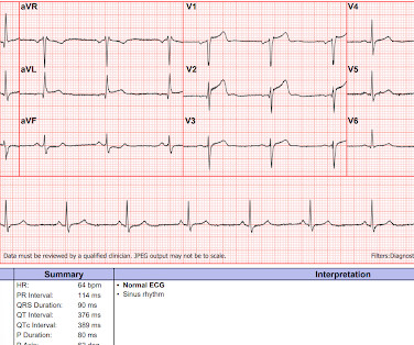
EMS arrived and recorded this ECG: What do you think? See same ECG below with computer automated interpretation, using the Glasgow ECG algorithm which apparently is used by many different providers and devices Amazing that the computer calls this normal. Next day ECG. And yet it still says "normal".
Written by Pendell Meyers I was reading ECGs in a database (without any clinical information) when I came to this one: What do you think? Seeing only this ECG with no context, I thought this ECG was within normal limits. So, if I had to interpret this ECG with no other context, I would say I see no clear evidence of OMI.
female, with a history of AF, presents complaining of palpitations intermittently over the last 4 days with a concurrent viral upper respiratory tract infection and this ECG
We organize all of the trending information in your field so you don't have to. Join 5,000+ users and stay up to date on the latest articles your peers are reading.
You know about us, now we want to get to know you!
Let's personalize your content
Let's get even more personalized
We recognize your account from another site in our network, please click 'Send Email' below to continue with verifying your account and setting a password.


Let's personalize your content