article thumbnail

ECG Blog #402 — Will Adenosine Convert This?

Ken Grauer, MD

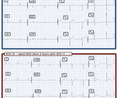
Figure-1: How would YOU interpret this ECG? MY Thoughts on the ECG in Figure-1: When faced with a challenging cardiac arrhythmia — It is a "luxury" to have access to a long lead rhythm strip containing 3 simultaneously -recorded leads. The repeat ECG after this treatment is shown in Figure-4.

EKG/ECG 419
article thumbnail

ECG Blog #405 — Is AV Block Complete (vs AV Dissociation)

Ken Grauer, MD

For full discussion of this case — See ECG Blog #191 — == The 2-lead rhythm strip shown in Figure-1 was obtained from an elderly woman who presented to the ED following a syncopal episode. ECG Media Pearl # 8 ( 8:20 minutes Video ) — ECG Blog #191 — Distinguishing between A V D issociation vs Complete AV Block ( 2/6/2021 ).

EKG/ECG 436
Insiders

Sign Up for our Newsletter

This site is protected by reCAPTCHA and the Google Privacy Policy and Terms of Service apply.

Trending Sources

article thumbnail

ECG Blog #411 — Is it Wenckebach?

Ken Grauer, MD

Figure-1: The initial ECG in today's case. KEY Clinical Point: If I was the medical provider charged with the care of the patient whose ECG is shown in Figure-1 — I would approach this tracing in the following sequential stages: I’d first establish that the patient was hemodnamically stable with this ECG and this cardiac rhythm.

EKG/ECG 441
article thumbnail

ECG Blog #415 — The Cath showed NO Occlusion!

Ken Grauer, MD

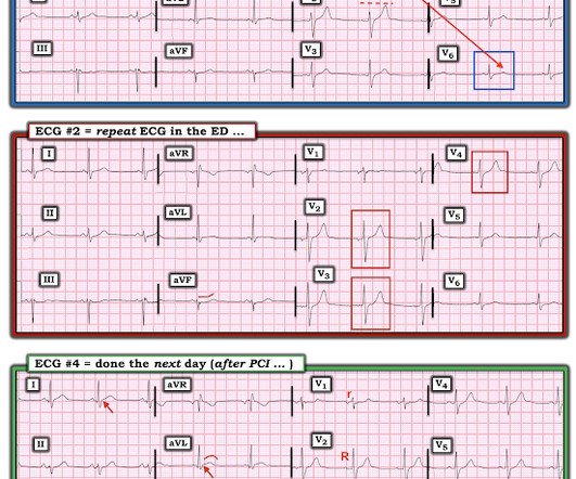
The ECG in Figure-1 was obtained following successful resuscitation. QUESTIONS: In view of the above history — How would YOU interpret the ECG in Figure-1 ? Is this ECG finding present in today’s initial ECG? Figure-1: The initial ECG in today's case — obtained after successful resuscitation from cardiac arrest. (

EKG/ECG 408
article thumbnail

ECG Blog #399 — Which Laddergram is Correct?

Ken Grauer, MD

The ECG in Figure-1 was obtained from a woman in her 60s — who was seen in the ED ( E mergency D epartment ) as part of her evaluation for trauma following a motor vehicle accident. Figure-1: The initial ECG in today's case. To do this — I apply the P s, Q s, 3 R Approach ( See ECG Blog #185 — for review of my system ).

EKG/ECG 397
article thumbnail

ECG Blog #393 — Why So Many Shapes?

Ken Grauer, MD

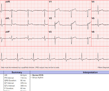
Figure-1: 12-lead ECG and long lead II rhythm strip obtained after Adenosine was given for a regular SVT rhythm. MY Thoughts on the ECG in Figure-1: As per the title of today's Blog post — there are numerous variations in the shape of the QRS complex over the course of the 17 beats seen in the long lead II rhythm strip.

EKG/ECG 285
article thumbnail

ECG Video Blog #404 (344) — Mobitz I, Mobitz II. or neither?

Ken Grauer, MD

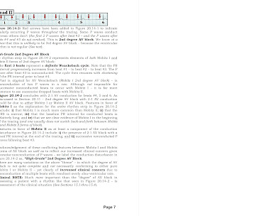
For full discussion of this case — See ECG Blog #344 — == How would YOU interpret the lead II rhythm strip shown in Figure-1 ? Section 2F ( 6 pages = the " short " Answer ) from my ECG-2014 Pocket Brain book provides quick written review of the AV Blocks ( This is a free download ). Or — Is it “ something else ”?

EKG/ECG 245