EM@3AM: Brainstem Strokes
EMDocs
MAY 11, 2024
Answer : Brainstem stroke specifically in the pons resulting in locked in syndrome. CT head without contrast 1 is performed and reveals the following: Question: What is the diagnosis?

EMDocs
MAY 11, 2024
Answer : Brainstem stroke specifically in the pons resulting in locked in syndrome. CT head without contrast 1 is performed and reveals the following: Question: What is the diagnosis?

RebelEM
FEBRUARY 24, 2025
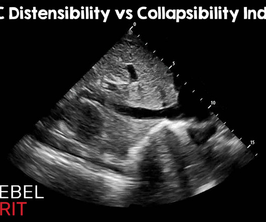
Of course, there are other methods of assessing fluid tolerance : Capillary refill evaluation, passive leg raise, central venous pressure measurement, pulmonary artery wedge pressures, stroke volume variation, pulse pressure variation, etc. Ultrasound Med Biol. Oct 2012; PMID: 23043910 Kumar A, et al. May 2014; PMID: 24495437.
This site is protected by reCAPTCHA and the Google Privacy Policy and Terms of Service apply.

ALiEM
JANUARY 29, 2024
It requires immediate consultation with ophthalmology as well as neurology as it is considered a stroke equivalent. Stroke workup for CRAO is necessary, and don’t forget about secondary prevention/risk stratification which must be part of the management. 2012 Dec;33(7):E263-E267. Epub 2012 Sep 21. Ultraschall Med.

RebelEM
MARCH 8, 2023
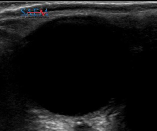
CRAO is a stroke of the eye; patients should be considered for a complete stroke work up. Signs Shafer’s sign (tobacco dust) May see a Weiss ring when the posterior vitreous (PV) detaches from the optic disc margin Visualization with ultrasound. Emergency Department Management: Emergency ophthalmology consultation. Read more

Pediatric Emergency Playbook
OCTOBER 1, 2017
Also used for severe MS, stroke, TBI, chronic pain. Ultrasound-guided refilling of an intrathecal baclofen pump—a case report. Are VNS safe in everyday life? Intrathecal Pumps Used to infuse basal rate of drug, usually baclofen for spasticity, but pump may contain morphine, bupivicaine, clonidine. Pediatr Neurosurg. 2013; 49:138–144.

Northwestern EM Blog
MAY 16, 2022
What is your initial imaging test of choice, ultrasound (US) or non-contrast CT, and why? Would you be confident in a point-of-care-ultrasound evaluation or a formal ultrasound? Many patients in the ultrasound groups did get additional imaging, but this was not the majority. How do you proceed? In this study, 40.7%

Taming the SRU
OCTOBER 7, 2024
history of VTE, increased calf circumference, pain and swelling), then the next step is to attain a compressible ultrasound of the affected leg. However, if clinical suspicion is high enough, doppler ultrasound, MR venography, or serial compressive ultrasound imaging can all be considered. Epub 2012 Oct 12. Haematologica.
Let's personalize your content