This site uses cookies to improve your experience. To help us insure we adhere to various privacy regulations, please select your country/region of residence. If you do not select a country, we will assume you are from the United States. Select your Cookie Settings or view our Privacy Policy and Terms of Use.
Cookie Settings
Cookies and similar technologies are used on this website for proper function of the website, for tracking performance analytics and for marketing purposes. We and some of our third-party providers may use cookie data for various purposes. Please review the cookie settings below and choose your preference.
Used for the proper function of the website
Used for monitoring website traffic and interactions
Cookie Settings
Cookies and similar technologies are used on this website for proper function of the website, for tracking performance analytics and for marketing purposes. We and some of our third-party providers may use cookie data for various purposes. Please review the cookie settings below and choose your preference.
Strictly Necessary: Used for the proper function of the website
Performance/Analytics: Used for monitoring website traffic and interactions
Shen 2013, Nickerson 2014, Scolaro 2016 ] Singh et al proposed an algorithm to guide treatment. Shen 2013, Nickerson 2014, Scolaro 2016 ] Singh et al proposed an algorithm to guide treatment. Shen 2013, Nickerson 2014, Scolaro 2016 ] Singh et al proposed an algorithm to guide treatment.
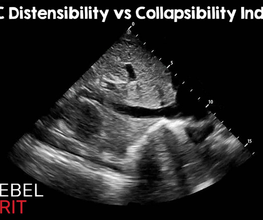
just be careful with pelvic fractures or any femoral arterial punctures/ devices. Regarding caval indexes, the advent of artificial intelligence and advanced learning has become integrated into many ultrasound machines. Ultrasound Med Biol. Oct 2012; PMID: 23043910 Kumar A, et al. May 2014; PMID: 24495437.
Bedside musculoskeletal ultrasound of his knees was performed. Credits: Andrew fried, md His left (normal) knee is shown below: Labeled combined still frames of the above ultrasound pointing out landmarks That should BE noted on every knee ultrasound. Sensation to light touch is intact distally.
You will notice the squeeze will cause no motion if there is a full rupture/tear, and diminished motion if there is a partial tear Performance Characteristics ( Garras 2012 ) Sensitivity Specificity (+) LR (-) LR 96-100% 93-100% 13.7 Partial rupture of the proximal Achilles tendon: a differential diagnostic problem in ultrasound imaging.
The current standard of practice has moved away from landmark-based central line placement given the efficacy and safety of ultrasound-based techniques. 2012 PMID: 21893125. This study also showed the median time for IO placement was only 1.2 minutes compared to a mean placement time of 10.7 minutes CVC group. Resuscitation.
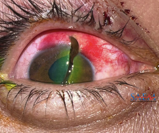
Laboratory Data CT Orbits/Sella w/ IV Contrast : No acute orbital fracture. Tonometry and ocular ultrasound (US) are generally not recommended as you could squeeze more liquid out of the eye or increase the intraocular pressure even more, pushing the iris further out. 2012 Feb;26(2):212-7. Visual acuity: able to count fingers.
EMS have already splinted an obvious mid-shaft femoral fracture, but he continues to be tachycardic and hypotensive. After a bedside ultrasound shows fluid in the right hemithorax, you insert an intercostal drain which immediately fills with one litre of blood. years ( 2 ).
3 Tenderness over the distal radial metaphysis after wrist injury is strongly suggestive of a distal radius fracture despite normal plain radiographs and fluoroscopic images. Children and older adults have weaker long bones than young adults and are more likely to sustain a distal radius fracture after a FOOSH than a carpal bone injury.
In fact, one of my surgeon grandfather's ortho buddies (perhaps with the help of some lunchtime martinis) took a look at the x-rays of my Boxer's Fracture and snapped it back into place without any analgesia or procedural sedation, casted me, and sent me home. EM makes a strong push into bedside use of ultrasound sonography.
Notably, lung ultrasound for the diagnosis of bacterial CAP demonstrated exceptional stand-alone diagnostic accuracy in 33 studies including 4,901 adults and children in the emergency department, with a pooled sensitivity of 92% and specificity of 90%. Raised inflammatory markers BMJ 2012; 344 :e454 doi:10.1136/bmj.e454 Gabay C, Kushner I.
Nachi: And if the prehospital team is lucky enough, or maybe unlucky enough, i don’t know, to have a credentialed provider who can perform ultrasound for those suspected of having a blunt cardiac injury, the general prehospital data on ultrasound is sparse. Jeff: Great, let’s move onto ED care, beginning with the H&P.
For example, a very anxious caregiver can easily transmit his or her anxiety to the child, which may either inhibit or amplify presentation of symptoms ( Bearden 2012 ). Long-bone injuries Fracture pain should be addressed immediately with splinting and analgesia. 2012 Jul;37(6):680-6. 2013 Dec 12;(12):CD004624. N Engl J Med.
23 Blunt carotid injury is more likely in patients with at least one of the following: Glasgow Coma Scale (GCS) ≤ 6, fracture of petrous bone, presence of diffuse axonal brain injury, and LeFort II/III fractures. 23 BCVI should be suspected with any high-energy trauma or any fractures of the maxilla or mandible. Tenenbaum, M.
Ankle fractures are the third most common fracture in the ED [2] and more than 20,000 patients are seen in the ED for ankle sprains each day [3]. traumatic axial loading with calcaneal fractures, pilons, and vertebral compression fractures) [5]. Gross deformities often suggest fracture or dislocation [Image 2].
Answer : Extensor tendon laceration Epidemiology: Hand extensor injuries make up >25% of orthopedic soft tissue injuries 2, 3 Common in young men in manual labor Dominant hand more likely to be injured 4 2012 study on 86 patients reported 83% men with a mean age of 34.2 Plast Reconstr Surg. 2013;132(4):560e-566e. doi:10.1097/PRS.0b013e3182a0148c
Upper limb fractures and dislocations can lead to associated arterial and nerve damage. First aid such as irrigation, tourniquet use, reduction and splinting of fractures/dislocations. Doppler Ultrasound: Non-invasive assessment of blood flow. Complex injuries including associated fractures or dislocations. Perkins, Z.
Potential causes include: -Trauma: Blunt or penetrating thoracic injuries, rib fractures, esophageal rupture (Boerhaave syndrome). Spontaneous pneumomediastinum on bedside ultrasound: case report and review of the literature. Pneumomediastinum Diagnosed on Ultrasound in the Emergency Department: A Case Report. West J Emerg Med.
We organize all of the trending information in your field so you don't have to. Join 5,000+ users and stay up to date on the latest articles your peers are reading.
You know about us, now we want to get to know you!
Let's personalize your content
Let's get even more personalized
We recognize your account from another site in our network, please click 'Send Email' below to continue with verifying your account and setting a password.


Let's personalize your content