Remove 2011 Remove Shock Remove Ultrasounds
article thumbnail

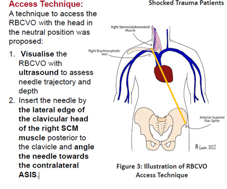
Is the blind Subclavian “trauma line” a thing of the past?

Greater Sydney Area HEMS

Obtaining access in shocked trauma patients can be notoriously difficult due to circulatory collapse. Those who are shocked, shut down with limited or no other options for peripheral access require central access. The evidence for improved safety and quality with the use of ultrasound for CVC implementation is well established [i].

article thumbnail

Approach to Shock

Pediatric Emergency Playbook

Do we recognize shock early enough? World wide, shock is a leading cause of morbidity and mortality in children, mostly for failure to recognize or to treat adequately. So, what is shock? Simply put, shock is the inadequate delivery of oxygen to your tissues. How do we prioritize our interventions? Pericardial Effusion?

Shock 40
Insiders

Sign Up for our Newsletter

This site is protected by reCAPTCHA and the Google Privacy Policy and Terms of Service apply.

Trending Sources

article thumbnail

Episode 28: LOST

PHEM Cast

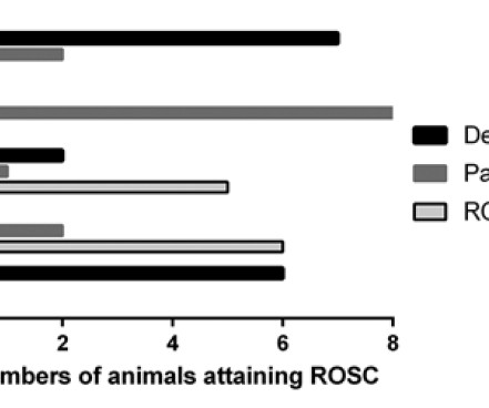
Ultrasound during TCA: Cureton et al. The heart of the matter: utility of ultrasound of cardiac activity during traumatic arrest. The outcomes from different resuscitative interventions in a haemorrhagic shock model in porcine model: From: Watts et al. Use of CPR in hemorrhagic shock, a dog model. Critical Care; 2011.

article thumbnail

REBEL Cast Ep123: Reduced-Dose Systemic Peripheral Alteplase in Massive PE?

RebelEM

Randomized, Controlled Trial of Ultrasound-Assisted Catheter-Directed Thrombolysis for Acute Intermediate-Risk Pulmonary Embolism. A prospective, Single-Arm Multicenter Trial of Ultrasound-Facilitated, Catheter-Directed, Low-Dose Fibrinolysis for Acute Massive and Submassive Pulmonary Embolism: The SEATTLE II Study. CHEST 2010.

Stroke 134
article thumbnail

POCUS findings of hemodynamically unstable PE with cardiac arrest

EMDocs

Point-of-Care-Ultrasound (POCUS) is a bedside modality that can assist Emergency Physicians (EPs) in differentiating PE from other causes of cardiac arrest. Multiorgan POCUS The diagnostic power of POCUS often resides in combining multiple ultrasound exams. 1-3 As many as 25% of acute PE cases present as sudden cardiac death.

article thumbnail

The Pediatric Surgical Abdomen

Pediatric Emergency Playbook

2011; 364(3):255-264. Hypertrophic pyloric stenosis: tips and tricks for ultrasound diagnosis. J Ultrasound. 2011; 29:1136-1140.:205-211. Necrotizing Enterocolitis. N Eng J Med. Niño DF et al. Necrotizing enterocolitis: new insights into pathogenesis and mechanisms. 2016; 13:590-600. Walsh MC et al. Semin Pedaitr Surg.

article thumbnail

What are treatment options for this rhythm, when all else fails?

Dr. Smith's ECG Blog

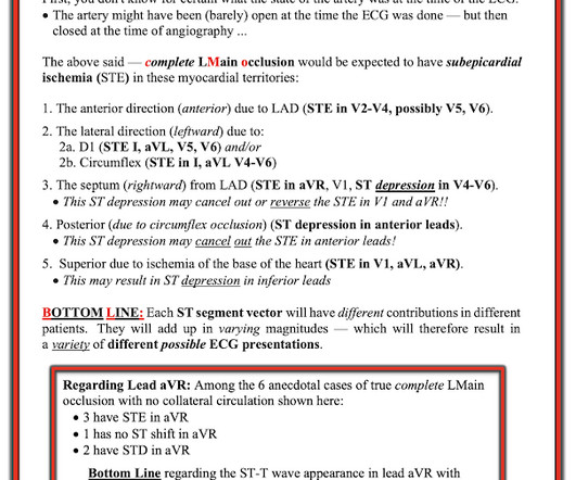
The patient in today’s case presented in cardiogenic shock from proximal LAD occlusion, in conjunction with a subtotally stenosed LMCA. Another approach is sympathetic chain (stellate ganglion) blockade if you have the skills to do it: it requires some expertise and ultrasound guidance. RCA — 100% proximal occlussion.

EKG/ECG 118