Remove 2011 Remove EKG/ECG Remove Wellness
article thumbnail

ECG Blog #422 — Was Clubbing an ECG Hint?

Ken Grauer, MD

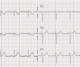
I was sent the ECG in Figure-1 — with the following history: The patient is a young man in his early 20s — who presents to the ED ( E mergency D epartment ) because of SOB ( S hortness O f B reath ) that had been ongoing for several hours. QUESTIONS: In view of the above history — How would YOU interpret the ECG in Figure-1 ?

EKG/ECG 468
article thumbnail

ECG Blog #384 — Why So Fast?

Ken Grauer, MD

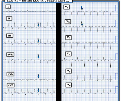
The ECG in Figure-1 was obtained from a young man with a history of rheumatic heart disease — who presented with “palpitations”. How would YOU interpret the ECG in Figure-1 ? Figure-1: The initial ECG in today’s case. To improve visualization — I've digitized the original ECG using PMcardio ). What is the rhythm?

EKG/ECG 365
Insiders

Sign Up for our Newsletter

This site is protected by reCAPTCHA and the Google Privacy Policy and Terms of Service apply.

Trending Sources

article thumbnail

ECG Blog #388 — Why Does Lead V1 Look Funny?

Ken Grauer, MD

The ECG in Figure-1 was obtained from an 18-year old woman — who moments before been resuscitated from out-of-hospital cardiac arrest. How would YOU interpret her post-resuscitation ECG? Does this ECG in Figure-1 provide clue(s) to the etiology of this patient's cardiac arrest? QUESTIONS: In light of the above clinical history.

EKG/ECG 370
article thumbnail

An undergraduate who is an EKG tech sees something. The computer calls it completely normal. How about the physicians?

Dr. Smith's ECG Blog

This was sent by an undergraduate (not yet in medical school, but applying now) who works as an ED technician (records all EKGs, helps with procedures, takes vital signs) and who reads this blog regularly. The undergraduate's analysis: This EKG shows J point elevation of about 0.5-1 Edited by Smith He also sent me this great case.

EKG/ECG 126
article thumbnail

ECG Blog #446 — What Kind of SVT?

Ken Grauer, MD

You are shown the ECG in Figure-1 — told only that the patient had a “continuous" tachycardia. QUESTIONS: How would YOU interpret the ECG in Figure-1 ? Figure-1: The initial ECG in today's case. ( To improve visualization — I've digitized the original ECG using PMcardio ). What is the differential diagnosis?

EKG/ECG 248
article thumbnail

ECG Blog #387 — 2 Minutes Later.

Ken Grauer, MD

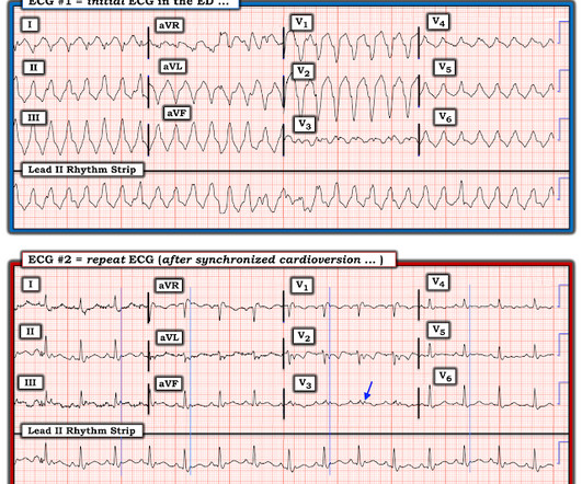
The ECG in Figure-1 was obtained from an elderly man with a history of coronary disease — who contacted EMS for "burning" chest discomfort that woke him at 3am. Some amount of time passed at home — during which his chest discomfort persisted The patient was hemodynamically stable at the time he was seen by EMS, when ECG #1 was recorded.

EKG/ECG 195
article thumbnail

ECG Blog #379 — Why Tachy on Telemetry?

Ken Grauer, MD

As discussed in detail in ECG Blog #240 — the term, “SVT” — refers to any supraventricular rhythm (ie, with origin at or above the AV node ) that manifests a rate of ≥100/minute. R elated E CG B log P osts to Today’s Case : ECG Blog #205 — Reviews my S ystematic A pproach to 12-lead ECG Interpretation.

EKG/ECG 195