Remove 2011 Remove EKG/ECG Remove Shock
article thumbnail

What are treatment options for this rhythm, when all else fails?

Dr. Smith's ECG Blog

The below ECG was recorded. The ECG shows obvious STEMI(+) OMI due to probable proximal LAD occlusion. This ECG does not have the typical ST-vector of an LAD occlusion. See below for Ken Grauer Comment on the initial ECG: == On arrival, another ECG was recorded: There appears to have been quite a bit of spontaneous reperfusion!

EKG/ECG 131
article thumbnail

ToxCard: Bupropion

EMDocs

Cardiogenic shock and hypotension can occur (systolic heart failure with reduced ejection fraction.) 10,12 Refractory shock or malignant arrhythmias occur in < 5% of patients. 3 Always get an EKG to assess for cardiotoxicity, including widened QRS and prolonged QTc, although this may not be evident right away. 2022; 55:232.e3-232.e4

Insiders

Sign Up for our Newsletter

This site is protected by reCAPTCHA and the Google Privacy Policy and Terms of Service apply.

Trending Sources

article thumbnail

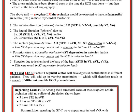
Diffuse Subendocardial Ischemia on the ECG. Left main? 3-vessel disease? No!

Dr. Smith's ECG Blog

An immediate 12-lead EKG was obtained: There is ST elevation in leads aVR and V1, with marked ST depression in I, II, III, aVF, V3-V6. DISCUSSION: The 12-lead EKG EMS initially obtained for this patient showed severe ischemia, with profound "infero-lateral" ST depression and reciprocal ST elevation in lead aVR. What should be done?

EKG/ECG 40
article thumbnail

POCUS findings of hemodynamically unstable PE with cardiac arrest

EMDocs

However, as shock resolved and hemodynamic stability improved, RV strain and underfilling of the LV remained apparent 30 minutes after ROSC (Vid 4). During a short period of ROSC an ECG was performed. The findings of RV strain on the ECG also supported the POCUS echo findings in this case. EKG RV strain. Circulation.

article thumbnail

Episode 7: Sepsis

PHEM Cast

Think about ECG as an example, So, where the box is green, the test has given us the correct result for the patient. One year mortality of patients treated with an emergency department based early goal directed therapy protocol for severe sepsis and septic shock: a before and after study. 2011 Aug 30;18(9):934–40. J Emerg Med.

Sepsis 52
article thumbnail

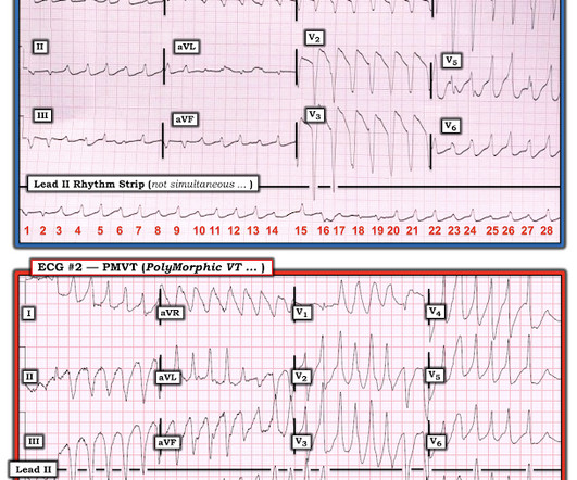
A Different Kind of Wide Rhythm -- Pleomorphic Ventricular Tachycardia

Dr. Smith's ECG Blog

== MY Comment by K EN G RAUER, MD ( 6/1 /2020 ): == YOU are asked to interpret the ECG shown in Figure-1. Figure-1: The initial ECG in the ED. MY THOUGHTS on ECG #1: My initial impression on looking at the ECG in Figure-1 — was that the rhythm was either rapid AFib in a patient with WPW — or — PMVT ( P oly M orphic VT ).

EKG/ECG 52
article thumbnail

MI in Children

Pediatric Emergency Playbook

Electrocardiography (ECG) should be performed on any patient with significant blunt chest injury. A negative ECG is highly consistent with no significant blunt myocardial injury. Any patient with a new abnormality on ECG (dysrhythmia, heart block, or signs of ischemia) should be admitted for continuous ECG monitoring.