Remove 2011 Remove EKG/ECG Remove Resuscitation
article thumbnail

ECG Blog #370 — A Post-Arrest Tachycardia.

Ken Grauer, MD

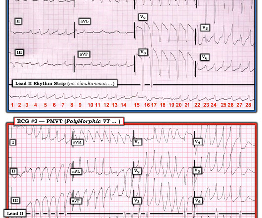
The 12-lead ECG and long lead II rhythm strip shown in Figure-1 — was obtained from a previously healthy, elderly woman who collapsed in the hospital parking lot. A series of cardiac arrhythmias were seen during the course of her resuscitation — including the interesting arrhythmia shown in the long lead II of Figure-1.

EKG/ECG 195
article thumbnail

A 40-something presented after attempted prehospital resuscitation with persistent Ventricular Fibrillation

Dr. Smith's ECG Blog

A 40-something with persistent Ventricular Fibrillation presented after attempted prehospital resuscitation A 40-something with no previous cardiac history presented to the ED in persistent Ventricular Fibrillation after attempted prehospital resuscitation. Finally, head-up CPR (which was not used here), makes for better resuscitation.

Insiders

Sign Up for our Newsletter

This site is protected by reCAPTCHA and the Google Privacy Policy and Terms of Service apply.

Trending Sources

article thumbnail

Diffuse Subendocardial Ischemia on the ECG. Left main? 3-vessel disease? No!

Dr. Smith's ECG Blog

An immediate 12-lead EKG was obtained: There is ST elevation in leads aVR and V1, with marked ST depression in I, II, III, aVF, V3-V6. In addition, the patient received 750 mL of fluid resuscitation with transient improvement of blood pressure. What should be done? Should the cath lab be activated?

EKG/ECG 40
article thumbnail

Episode 7: Sepsis

PHEM Cast

It is worth noting, that with “Sepsis 3” many of these terms will become out-of-date – but validation work is required… The Rivers’ paper can be accessed here: [link] It was a single centre study which compared standard care with protocolised resuscitation packaged together as early goal-directed therapy (EGDT).

Sepsis 52
article thumbnail

A Different Kind of Wide Rhythm -- Pleomorphic Ventricular Tachycardia

Dr. Smith's ECG Blog

== MY Comment by K EN G RAUER, MD ( 6/1 /2020 ): == YOU are asked to interpret the ECG shown in Figure-1. Figure-1: The initial ECG in the ED. MY THOUGHTS on ECG #1: My initial impression on looking at the ECG in Figure-1 — was that the rhythm was either rapid AFib in a patient with WPW — or — PMVT ( P oly M orphic VT ).

EKG/ECG 52
article thumbnail

ECG Blog #388 — Why Does Lead V1 Look Funny?

Ken Grauer, MD

The ECG in Figure-1 was obtained from an 18-year old woman — who moments before been resuscitated from out-of-hospital cardiac arrest. How would YOU interpret her post-resuscitation ECG? Does this ECG in Figure-1 provide clue(s) to the etiology of this patient's cardiac arrest? About A RVC/ A RVD.

EKG/ECG 370
article thumbnail

ECG Blog #436 — Bigeminy or Alternans?

Ken Grauer, MD

The ECG in Figure-1 — was obtained from an older man with known coronary disease. He developed cardiac arrest shortly after the ECG in Figure-1 was recorded. QUESTIONS: How would YOU interpret the ECG in Figure-1 ? QUESTIONS: How would YOU interpret the ECG in Figure-1 ? Figure-1: The initial ECG in today's case. (

EKG/ECG 399