Remove 2008 Remove Documentation/Coding Remove EKG/ECG
article thumbnail

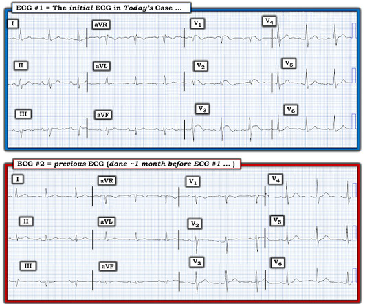
A man in his 30s with chest pain. How was he managed? What if they had used the Queen of Hearts?

Dr. Smith's ECG Blog

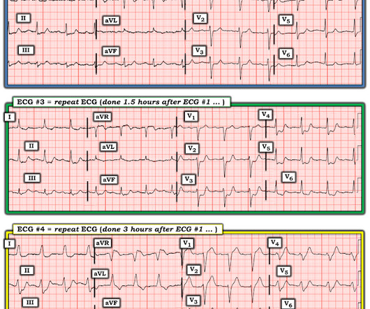
Triage ECG: And here she explains her assessment: The ECG was read as simply "No ST elevation." No repeat ECG was done at this time. Repeat ECG shows no changes." Here is that repeat ECG below, around 3 hours after triage: Repeat troponin during delay rose to 18,700 ng/L. Which is true. None further were ordered.

EKG/ECG 121
article thumbnail

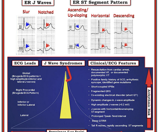
Acute OMI or "Benign" Early Repolarization?

Dr. Smith's ECG Blog

The following ECG was obtained. Note that the machine read is "normal sinus rhythm, normal ECG." ECG 1 What do you think? I sent this ECG to Dr. Smith and Dr. Meyers with no clinical context. Smith comment: this troponin alone should be enough data to activate the cath lab, regardless of the ECG. <0.049 ng/mL).

EKG/ECG 131
Insiders

Sign Up for our Newsletter

This site is protected by reCAPTCHA and the Google Privacy Policy and Terms of Service apply.

Trending Sources

article thumbnail

A man in his 70s with weakness and syncope

Dr. Smith's ECG Blog

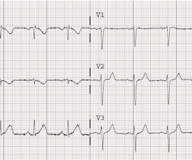
An ECG was performed in the ED at 1554: Original image unavailable, this is the only recorded scanned ECG available. In a patient with syncope and fever, this ECG looks more like Brugada. Smith comment: the ECG in question could be due to Brugada, even though there is a change from baseline. PM Cardio digitized version.

EKG/ECG 92
article thumbnail

See this "NSTEMI" go unrecognized for what it really is, how it progresses, and what happens

Dr. Smith's ECG Blog

The baseline ECG is basically normal with no ischemia. Here is what the Queen of Hearts says about the Baseline ECG: Active chest pain triage ECG also read as not OMI: She says "not OMI", but she does not have access to the baseline ECG. In the future, we will be able to have her compare with previous and serial ECGs.

EKG/ECG 83
article thumbnail

Compare these two ECGs. Do either, neither, or both show anything important?

Dr. Smith's ECG Blog

Sean Rees MD, written by Pendell Meyers, other case by Sam Ghali and Steve Smith Take a look at these two ECGs below from two patients in the ED, first without any clinical context. The Queen of Hearts correctly says: Smith : Why is this ECG which manifests so much ST Elevation NOT a STEMI (even if it were a 60 year old with chest pain)?

EKG/ECG 52
article thumbnail

Hyperthermia and ST Elevation

Dr. Smith's ECG Blog

An initial EKG was obtained: Computer read: sinus tachycardia, early acute anterior infarct. Here is her prior EKG: When compared to the old EKG – Q waves present before, TWI in aVR present before, but all other changes are new. What is the differential for this EKG? T wave inversion III, aVR, TWF in aVF. Is this an OMI?

article thumbnail

Inferior Hyperacute T-waves

Dr. Smith's ECG Blog

He called 911 and paramedics recorded a prehospital 12 lead ECG which showed a clear inferior STEMI (not shown, tracing could not be found). On arrival, the following ECG was recorded. In this case the diagnosis was clear due to the prehospital ECG. But suppose there had been no prehospital ECG? There is 0.5

EKG/ECG 52