article thumbnail

Case Report: The Not So Normal, Normal EKG

ACEP Now

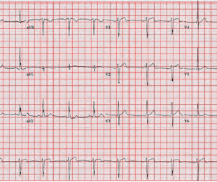
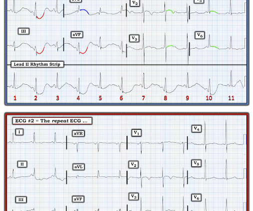
The patient had an EKG performed within 10 minutes of arrival while in triage (see Figure 1). In the absence of significant ST elevations, the EKG was signed and the patient was placed back in the queue to await a bed. FIGURE 1: First ED EKG. Given his EKG findings and presentation, he was started on heparin and admitted.

EKG/ECG 52
article thumbnail

Acute artery occlusion -- which one?

Dr. Smith's ECG Blog

Her ECG is shown below: What do you think? The conventional machine algorithm interpreted this ECG as STEMI. Alternatively, with STE in V1 and III, and STD in I and aVL, this ECG could represent proximal RCA OMI with right ventricular involvement. What do you do clinically when the ECG looks like this?

EKG/ECG 126
Insiders

Sign Up for our Newsletter

This site is protected by reCAPTCHA and the Google Privacy Policy and Terms of Service apply.

Trending Sources

article thumbnail

Does this T wave pattern mean anything?

Dr. Smith's ECG Blog

An Initial ECG was performed: Initial ECG: Sinus tachycardia with prolonged QT interval (QTc of 534 ms by Bazett). A repeat ECG was performed 2 hours after arrival: QTc prolongation ato 722 ms now with alternating T wave pattern (T wave alternans) I texted this to Smith who responded: “T wave alternans and long QT. Narayan SM.

EKG/ECG 130
article thumbnail

ACMT Toxicology Visual Pearl: Salt, not Shock

ALiEM

What agent would most likely be responsible for these ECG findings? This EKG shows a wide complex (QRS 240 msec), irregular rhythm with left bundle branch block morphology at a rate slower than expected (90 bpm) for a ventricular arrhythmia such as ventricular tachycardia. 2007 Apr;19(2):155-9. PMID: 35287973. doi: 10.1111/j.1742-6723.2006.00909.x.

Shock 73
article thumbnail

I was shown this ECG without any information. What do you think?

Dr. Smith's ECG Blog

I was shown this ECG; "Steve, what do you think of this?": Whenever you see a very bizarre EKG, you should think about pulse tapping artifact. Lead I in this ECG is not bizarre [though it does show a very long QT (or QU)]. Solution : repeat the ECG, but move the left leg electrode. At 44 minutes, another ECG was recorded.

EKG/ECG 52
article thumbnail

Acute dyspnea in an older woman, is it OMI?

Dr. Smith's ECG Blog

ECG is shown below. Many ECG readers will not comment any further on rhythm once ventricular pacing has been identified, but it is still critical to determine the atrial rhythm. Cardiology felt that there was baseline artifact and recommended immediate repeat ECG which is shown below. What do you think?

EKG/ECG 108
article thumbnail

Putting Clinical Gestalt to Work in the Emergency Department

ACEP Now

Does that normal troponin and ECG obviate the need for cardiology consultation for my patient with a concerning story for acute coronary syndrome? The “bundling” heuristic—if I send a troponin, then an ECG is needed. Should I draw a venous or arterial blood gas before deciding on intubation? Mary’s Hospital in Leonardtown, Maryland.