Remove 2005 Remove EKG/ECG Remove Ultrasounds
article thumbnail

Chest Pain in Children: ReBaked Morsel

Pediatric EM Morsels

EKG Reasonable screen for cardiac etiology [ Kane, 2010 ]: Chest Pain with Exertion? Ultrasound diagnosis of occult pneumothorax. 2005 Jun;33(6):1231-8. The role of point-of-care ultrasound in the diagnosis of pericardial effusion: a single academic center retrospective study. Ultrasound J. Crit Care Med.

EKG/ECG 271
article thumbnail

46 year old with chest pain develops a wide complex rhythm -- see many examples

Dr. Smith's ECG Blog

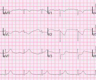
These diagnoses were not found in his medical records nor even a baseline ECG. An ECG was obtained shortly after arrival: What do you think? There is no evidence of WPW on this ECG, but it is diagnostic for OMI. He reported a history of “Wolf-Parkinson-White” and “heart attack” but said neither had been treated.

EKG/ECG 107
Insiders

Sign Up for our Newsletter

This site is protected by reCAPTCHA and the Google Privacy Policy and Terms of Service apply.

Trending Sources

article thumbnail

Severe shock, obtunded, and a diagnostic prehospital ECG. Also: How did this happen?

Dr. Smith's ECG Blog

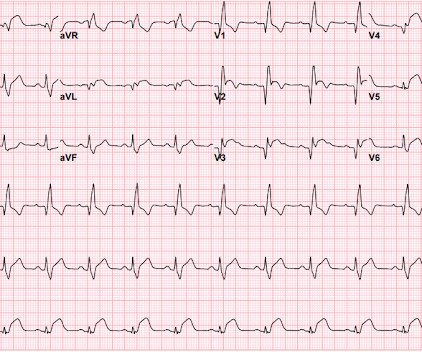
A prehospital ECG was recorded: Limb leads: Precordial Leads What is the therapy? On arrival, the patient was in shock, was intubated, and had an immediate cardiac ultrasound. What does a heart look like on ultrasound when the EKG looks like that? The followup ECG is here: Now the QRS is only slightly prolonged.

Shock 40
article thumbnail

A teenager with chest pain, a troponin below the limit of detection, and "benign early repolarization"

Dr. Smith's ECG Blog

Meyers ECG Interpretation: Easily diagnostic of LAD occlusion. The ECG easily meets STEMI criteria in all leads V2-V6, as well. I sent this ECG to Dr. Smith, with the only information that it is a 17 year old with chest pain. The ECG was interpreted as "benign early repolarization." Beware a negative Bedside ultrasound.

EKG/ECG 52
article thumbnail

STEMI with Life-Threatening Hypokalemia and Incessant Torsades de Pointes

Dr. Smith's ECG Blog

Bedside ultrasound showed no effusion and moderately decreased LV function, with B-lines of pulmonary edema. Here is his ED ECG: There is obvious infero-posterior STEMI. This subsequent ECG was recorded after the K was up to 2.2 These two rhythms are often indistinguishable on the monitor or ECG. This was stented.

article thumbnail

MI in Children

Pediatric Emergency Playbook

Electrocardiography (ECG) should be performed on any patient with significant blunt chest injury. A negative ECG is highly consistent with no significant blunt myocardial injury. Any patient with a new abnormality on ECG (dysrhythmia, heart block, or signs of ischemia) should be admitted for continuous ECG monitoring.

article thumbnail

Emergency Department Syncope Workup: After H and P, ECG is the Only Test Required for Every Patient.

Dr. Smith's ECG Blog

Check : [vitals, SOB, Chest Pain, Ultrasound] If the patient has Abdominal Pain, Chest Pain, Dyspnea or Hypoxemia, Headache, Hypotension , then these should be considered the primary chief complaint (not syncope). Abnormal ECG – looks for cardiac syncope. Aortic Dissection, Valvular (especially Aortic Stenosis), Tamponade.