This site uses cookies to improve your experience. To help us insure we adhere to various privacy regulations, please select your country/region of residence. If you do not select a country, we will assume you are from the United States. Select your Cookie Settings or view our Privacy Policy and Terms of Use.
Cookie Settings
Cookies and similar technologies are used on this website for proper function of the website, for tracking performance analytics and for marketing purposes. We and some of our third-party providers may use cookie data for various purposes. Please review the cookie settings below and choose your preference.
Used for the proper function of the website
Used for monitoring website traffic and interactions
Cookie Settings
Cookies and similar technologies are used on this website for proper function of the website, for tracking performance analytics and for marketing purposes. We and some of our third-party providers may use cookie data for various purposes. Please review the cookie settings below and choose your preference.
Strictly Necessary: Used for the proper function of the website
Performance/Analytics: Used for monitoring website traffic and interactions
A 12-lead EKG shows sinus tachycardia but is otherwise normal. Both can result in heat exhaustion and heat stroke and have many overlapping symptoms. Patients with heat stroke have hot, dry skin and altered mental status (e.g., C, and heat stroke occurs at a core temperature > 40°C. Temps greater than 41.5C
Electrocardiography (ECG) should be performed on any patient with significant blunt chest injury. A negative ECG is highly consistent with no significant blunt myocardial injury. Any patient with a new abnormality on ECG (dysrhythmia, heart block, or signs of ischemia) should be admitted for continuous ECG monitoring.
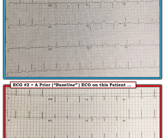
This ECG was recorded on arrival: What do you think? Proof that all STE and hyperacute T-waves in the presentation ECG are new. They collected several repeat ECGs at the outside hospital before transport: None of these three ECGs meet STEMI criteria. This ECG shows persistent Occlusion MI but does not meet STEMI criteria.
Abnormal ECG – looks for cardiac syncope. Abnormal Electrocardiogram (ECG): Defined (San Fran syncope rule) as any new changes when compared to the last ECG or presence of non-sinus rhythm. If no previous ECG was available, ECG was classified as abnormal if any abnormality was present. orthostatic vitals b.
Pain can be associated with a friction rub on cardiac auscultation, a pericardial effusion on a bedside echocardiogram, or diffuse ST elevations on an EKG. 2005 Jul-Aug 2005;12(4):311-9. Up to two-thirds of rib fractures are missed on initial chest radiographs. Jan 23 2008;(1):CD000396. doi:10.1002/14651858.CD000396.pub3
We organize all of the trending information in your field so you don't have to. Join 5,000+ users and stay up to date on the latest articles your peers are reading.
You know about us, now we want to get to know you!
Let's personalize your content
Let's get even more personalized
We recognize your account from another site in our network, please click 'Send Email' below to continue with verifying your account and setting a password.


Let's personalize your content