Remove 2005 Remove EKG/ECG Remove Resuscitation
article thumbnail

ECG Blog #448 — A Young Man with Chest Pain.

Ken Grauer, MD

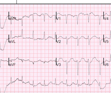
The ECG in Figure-1 was obtained from a previously healthy man in his early 20s — who initially presented with GI symptoms, that then evolved into CP ( C hest P ain ). QUESTIONS: Given the above history — How would YOU interpret the initial ECG that is shown in Figure-1 ? Figure-1: The initial ECG in today's case. (

EKG/ECG 296
article thumbnail

Patient with severe DKA, look at the ECG

Dr. Smith's ECG Blog

Here is the ECG: Sinus tachycardia. This is an extremely dangerous ECG. When the ECG shows the effects of hypokalemia, it is particularly dangerous. Here are the American Heart Association Guidelines: 2005 American Heart Association Guidelines for Cardiopulmonary Resuscitation and Emergency Cardiovascular Care Part 10.1:

EKG/ECG 52
Insiders

Sign Up for our Newsletter

This site is protected by reCAPTCHA and the Google Privacy Policy and Terms of Service apply.

Trending Sources

article thumbnail

Severe shock, obtunded, and a diagnostic prehospital ECG. Also: How did this happen?

Dr. Smith's ECG Blog

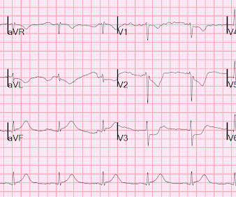
A prehospital ECG was recorded: Limb leads: Precordial Leads What is the therapy? What does a heart look like on ultrasound when the EKG looks like that? The followup ECG is here: Now the QRS is only slightly prolonged. A middle-aged woman was found down in her apartment unconscious. She was in shock with thready pulses.

Shock 40
article thumbnail

Prehospital Traumatic Cardiac Arrest: A Systematic Review and Meta-analysis

RebelEM

The only statistically significant factor intra-arrest was the first ECG rhythm, which only had an odds ratio of 1.12, which is relatively low. However, we might expect on-scene advanced trauma resuscitation to prolong prehospital time sizably. Cardiopulmonary resuscitation traumatic cardiac arrest—there are survivors.

article thumbnail

STEMI with Life-Threatening Hypokalemia and Incessant Torsades de Pointes

Dr. Smith's ECG Blog

Here is his ED ECG: There is obvious infero-posterior STEMI. After resuscitation, he was found to have a 90% thrombotic lesion in the same saphenous vein graft to the right posterior descending artery. This subsequent ECG was recorded after the K was up to 2.2 These two rhythms are often indistinguishable on the monitor or ECG.

article thumbnail

MI in Children

Pediatric Emergency Playbook

Electrocardiography (ECG) should be performed on any patient with significant blunt chest injury. A negative ECG is highly consistent with no significant blunt myocardial injury. Any patient with a new abnormality on ECG (dysrhythmia, heart block, or signs of ischemia) should be admitted for continuous ECG monitoring.

article thumbnail

EM@3AM: Hyperthermia

EMDocs

A 12-lead EKG shows sinus tachycardia but is otherwise normal. Evaporative cooling is the preferred method to actively reduce body temperature in the emergency department , as it can be performed with ongoing resuscitation efforts. Resuscitation. , 148 , 173–190. Temps greater than 41.5C Temps greater than 41.5C Epstein, J.,