article thumbnail

ECG Blog #448 — A Young Man with Chest Pain.

Ken Grauer, MD

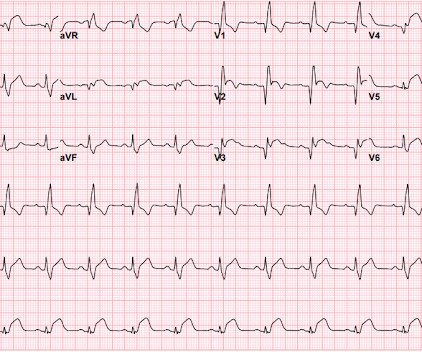
The ECG in Figure-1 was obtained from a previously healthy man in his early 20s — who initially presented with GI symptoms, that then evolved into CP ( C hest P ain ). QUESTIONS: Given the above history — How would YOU interpret the initial ECG that is shown in Figure-1 ? Figure-1: The initial ECG in today's case. (

EKG/ECG 296
article thumbnail

Chest Pain in Children: ReBaked Morsel

Pediatric EM Morsels

EKG Reasonable screen for cardiac etiology [ Kane, 2010 ]: Chest Pain with Exertion? 2005 Jun;33(6):1231-8. Is there a family history of sudden death, hypertrophic cardiomyopathy, long QT syndrome, or tachyarrhythmias? Chest Pain in Children: A Reasonable Work-up The goal is to balance risk of the rare with risk of over-testing.

EKG/ECG 271
Insiders

Sign Up for our Newsletter

This site is protected by reCAPTCHA and the Google Privacy Policy and Terms of Service apply.

Trending Sources

article thumbnail

Texted from a former EM resident: 70 yo with syncope and hypotension, but no chest pain. Make their eyes roll!

Dr. Smith's ECG Blog

Here is the ED ECG (a photo of the paper printout) What do you think? American Journal of Emergency Medicine 2005; 23(3):279-287. The "good news" — is that a cardiac-related cause of syncope is unlikely if the initial ECG is normal, and cardiac monitoring in the ED fails to reveal significant arrhythmia.

EMS 114
article thumbnail

46 year old with chest pain develops a wide complex rhythm -- see many examples

Dr. Smith's ECG Blog

These diagnoses were not found in his medical records nor even a baseline ECG. An ECG was obtained shortly after arrival: What do you think? There is no evidence of WPW on this ECG, but it is diagnostic for OMI. If the LAD is completely occluded, then why does the ECG show reperfusion? What are we seeing here?

EKG/ECG 107
article thumbnail

Patient with severe DKA, look at the ECG

Dr. Smith's ECG Blog

Here is the ECG: Sinus tachycardia. This is an extremely dangerous ECG. When the ECG shows the effects of hypokalemia, it is particularly dangerous. Here are the American Heart Association Guidelines: 2005 American Heart Association Guidelines for Cardiopulmonary Resuscitation and Emergency Cardiovascular Care Part 10.1:

EKG/ECG 52
article thumbnail

Classic LV aneurysm (persistent ST elevation after previous MI)

Dr. Smith's ECG Blog

What are the worrisome EKG findings, and what is the differential diagnosis? If you want a rule, I derived one a few years ago (AJEM 23(3):279-287; May 2005). I cannot tell from this ECG if it is 12 hours or 12 years since the MI. 70 yo with h/o MI, recent CABG, has acute onset of chest pain. What is the most likely diagnosis?

EKG/ECG 52
article thumbnail

Severe shock, obtunded, and a diagnostic prehospital ECG. Also: How did this happen?

Dr. Smith's ECG Blog

A prehospital ECG was recorded: Limb leads: Precordial Leads What is the therapy? What does a heart look like on ultrasound when the EKG looks like that? The followup ECG is here: Now the QRS is only slightly prolonged. A middle-aged woman was found down in her apartment unconscious. She was in shock with thready pulses.

Shock 40