article thumbnail

ECG Blog #380 — What is "Swirl"?

Ken Grauer, MD

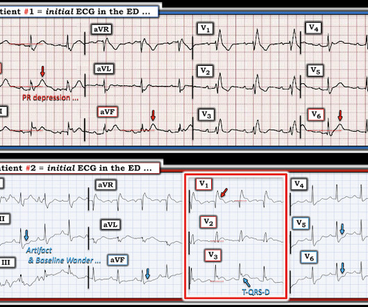
The ECG in Figure-1 — was obtained from an older woman with persistent CP ( C hest P ain ) over the previous day. Figure-1: The initial ECG in today's case. Voltage for LVH is satisfied — at least by Peguero Criteria ( Sum of deepest S in any chest lead + S in V4 ≥23 mm in a woman — as discussed in ECG Blog #73 ).

EKG/ECG 363
article thumbnail

SAEM Clinical Images Series: A Rare Pediatric Scalp Rash

ALiEM

In addition, an EKG is essential given that neonates can present with irreversible total atrioventricular heart block, which can present in utero or after birth. Any baby with findings concerning for neonatal lupus should have an EKG performed. 2004 Jan;9(1):52-6. References Diaz-Frias J, Badri T. Neonatal Lupus Erythematosus.

Insiders

Sign Up for our Newsletter

This site is protected by reCAPTCHA and the Google Privacy Policy and Terms of Service apply.

Trending Sources

article thumbnail

A Tough ECG, But Learn From It!

Dr. Smith's ECG Blog

I want all to know that, with the right mind preparation, and the use of the early repol/LAD occlusion formula, extremely subtle coronary occlusion can be detected prospectively, with no other information than the ECG. His ECG was repeated at this point: This shows a well developed anterior STEMI. His first troponin was normal.

EKG/ECG 52
article thumbnail

What does the angiogram show? The Echo? The CT coronary angiogram? How do you explain this?

Dr. Smith's ECG Blog

She has no previous cardiac history of which she is aware 911 was called and here is her Prehospital ECG: What do you think? link] Case continued She arrived in the ED and here is the first ED ECG. In comparison with the first ECG, I would guess that the artery is reperfusing. N Engl J Med [Internet] 2013;368(21):2004–13.

EKG/ECG 109
article thumbnail

Two patients with chest pain and RBBB: do either have occlusion MI?

Dr. Smith's ECG Blog

Past medical history included RBBB without other cardiac history, but old ECG was not available. As for the ECG, it could represent OMI, but RBBB is also a clue that it may be PE. But with prehospital and ED ECGs being ‘STEMI negative’, the ECG was signed off and the patient waited to be seen.

EKG/ECG 72
article thumbnail

Upon arrival to the emergency department, a senior emergency physician looked at the ECG and said "Nothing too exciting."

Dr. Smith's ECG Blog

Our first 12 lead ECG was then recorded at 0926. Another ECG was recorded 5 minutes later just before arrival at the hospital: Similar The patient was transported to a nearby suburban hospital with PCI capabilities while my partner cared for her. Smith: this ECG and clinical presentation is diagnostic of LAD Occlusion.

EKG/ECG 52
article thumbnail

Wide-complex tachycardia that didn’t follow the rules

Dr. Smith's ECG Blog

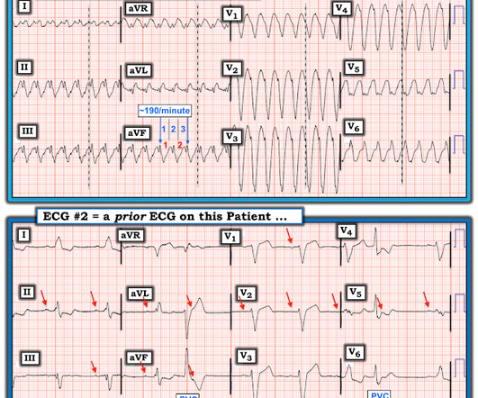
Case contributed by Brooks Walsh , an emergency physician and ECG aficionado in Connecticut (and a few comments by Smith) A middle-aged patient came to the ED complaining of palpitations, dyspnea, sweating, and chest pressure that radiated to the arms. Initial ECG in the ED: Presenting ECG : Wide-complex tachycardia at a rate about 200.

EKG/ECG 52