article thumbnail

A teenager involved in a motor vehicle collision with abnormal ECG

Dr. Smith's ECG Blog

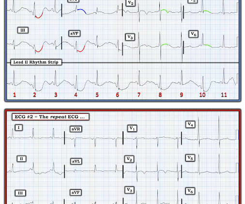
Here is his initial ECG around 1330: What do you think? The ECG shows sinus tachycardia with RBBB and LAFB, without clear additional superimposed signs of ischemia. Initial high sensitivity troponin I: 3,830 ng/L (URL 20 ng/L for men) 1445: Similar to initial ECG. He was intubated for altered mental status.

EKG/ECG 116
article thumbnail

emDOCs Revamp: Alcohol Withdrawal

EMDocs

2003; 348:1786-1795. fold higher risk of NSTI than the control group 12 For those without comorbidities , AUD exhibited a 15.2-fold Management of drug and alcohol withdrawal. N Engl JMed. Gortney J, Raub J, Patel P, et al. Alcohol withdrawal syndrome in medical patients. Cleve Clin J Med. 2016; 83(1): 67-79. Muncie H, Yasinian Y, Oge L.

Seizures 104
Insiders

Sign Up for our Newsletter

This site is protected by reCAPTCHA and the Google Privacy Policy and Terms of Service apply.

article thumbnail

SGEM#430: De Do Do Do, De Dash, Dash DAShED – Diagnosing Acute Aortic Syndrome in the ED.

The Skeptics' Guide to EM

The ECG shows sinus tachycardia with non-specific ST-T changes. This study found that between 2003 and 2018, there were 1,299 cases of AAS in Ontario, the largest province in the country. She is bradycardic, hypotensive, and tachypneic. She is received in the resuscitation room. The chest x-ray is unremarkable. per 100,000. [4-8]

article thumbnail

ACMT Toxicology Visual Pearl: The Heart Won’t Go On and On

ALiEM

An ECG may show signs similar to digoxin exposure Classic “scooping” of ST segments Heart blocks Dysrhythmias Hyperkalemia may be seen with severe cardiac glycoside poisoning. Serial ECGs and monitoring of serum potassium should be done in cases of severe poisoning. Lancet 2003; 361 (9373): 1935-8.

article thumbnail

Patient in Single Vehicle Crash: What is this ST Elevation, with Peak Troponin of 6500 ng/L?

Dr. Smith's ECG Blog

As a routine part of his critical trauma evaluation, he had an ECG recorded: There is an rSR" in V1 and V2, with downsloping ST segment and inverted T-wave which is very similar to a Brugada Type 1 phenocopy. On the other hand, the RV lies directly under leads V1 and V2; this makes pneumothorax less likely as an etiology of the abnormal ECG.

EKG/ECG 52
article thumbnail

Diffuse Subendocardial Ischemia on the ECG. Left main? 3-vessel disease? No!

Dr. Smith's ECG Blog

An immediate 12-lead EKG was obtained: There is ST elevation in leads aVR and V1, with marked ST depression in I, II, III, aVF, V3-V6. DISCUSSION: The 12-lead EKG EMS initially obtained for this patient showed severe ischemia, with profound "infero-lateral" ST depression and reciprocal ST elevation in lead aVR. What should be done?

EKG/ECG 40
article thumbnail

What is strange about this paroxysmal atrial fibrillation in an otherwise healthy patient? And what happened after giving ibutilide?

Dr. Smith's ECG Blog

Here is her EKG: What is unusual about this? Here is the ECG after ibutilide: What do you notice? Here is the post-cardioversion ECG: Sinus rhythm, still with the longer QT interval. For more on SSS — See My Comment at the bottom of the page in the July 5, 2018 post in Dr. Smith’s ECG Blog. She was on no medications.

EKG/ECG 52