article thumbnail

Very fast regular tachycardia: 2 ECGs from the same patient. What is going on?

Dr. Smith's ECG Blog

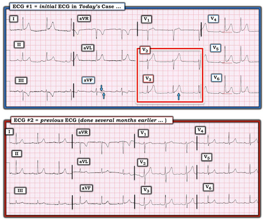
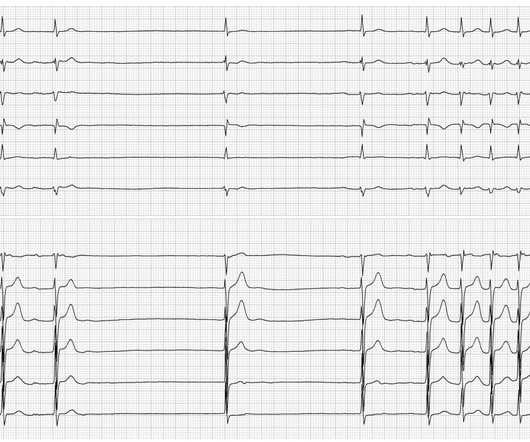
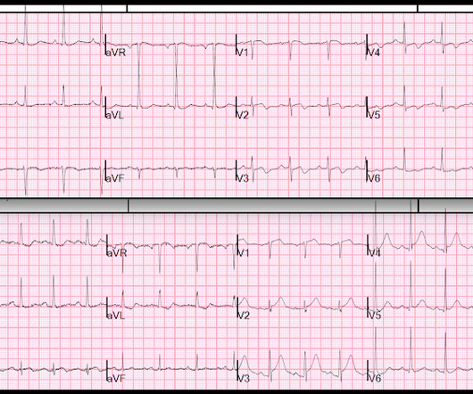
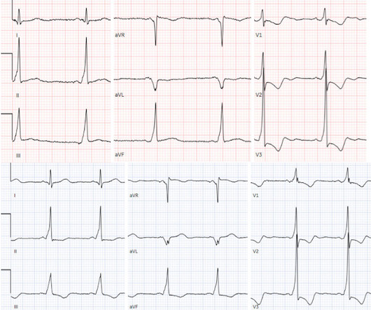
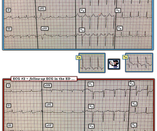
An ECG was recorded immediately and is shown below. How do you interpret the ECG? ECG#1 There is a regular tachycardia with a ventricular rate of about 180 bpm. After cardioversion, if successful, you can take a few moments to assess the 12-lead in more detail and assess the post conversion ECG. ECG#2 What do you think?

EKG/ECG 133
article thumbnail

An elderly male with shortness of breath

Dr. Smith's ECG Blog

ECG 1 at time zero EARLY REPOLARIZATION ABNORMAL ECG ED final official overread: "early repol vs hyperacute T, minimal changes from previous (previous shown below)" What do YOU think? Thus, the LAD has reperfused ("recanalized") spontaneously Queen on ECG 2: Not OMI, Low confidence She is also worried about V2.

EKG/ECG 116
Insiders

Sign Up for our Newsletter

This site is protected by reCAPTCHA and the Google Privacy Policy and Terms of Service apply.

Trending Sources

article thumbnail

SSS

ECG Guru

The ECG shows an example of a patient with bradycardia/tachycardia syndrome. After a further pause of just under 2000 ms, 2 sinus node beats follow, which merge into a sinus tachycardia or atrial tachycardia (heart rate approx. Initially, a sinus rhythm with a heart rate of approx. 70 bpm is seen. 120 bpm here).

EKG/ECG 75
article thumbnail

First ED ECG is Wellens' (pain free). What do you think the prehospital ECG showed (with pain)?

Dr. Smith's ECG Blog

He arrived in the ED and had this ECG recorded:  There are Wellens' waves, type A (upsloping ST segment then inversion of the terminal part of the T-wave - terminal T-wave inversion, or biphasic T-waves) in V2-V4, and aVL. When the patient had chest pain, prior to nitroglycerine, what do you think the ECG showed ? A stent was placed.

EKG/ECG 52
article thumbnail

Sick Sinus Syndrome

ECG Guru

The ECG shows an example of a patient with bradycardia/tachycardia syndrome (also called sick sinus syndrome). After a further pause of just under 2000 ms, 2 sinus node beats follow, which merge into a sinus tachycardia or atrial tachycardia (heart rate approx. Initially, a sinus rhythm with a heart rate of approx. 70 bpm is seen.

EKG/ECG 52
article thumbnail

A woman in her 50s with chest pain and dyspnea

Dr. Smith's ECG Blog

Here is her triage ECG: PM Cardio Version (see original screenshot I received below) Original image. Here is the prior ECG on file (from 1 month ago, when she was having "NSTEMI" with high sensitivity troponin peaking at 200 ng/L): What do you think now that you can see the prior ECG? Her vitals were within normal limits.

EKG/ECG 52
article thumbnail

Two patients with chest pain and RBBB: do either have occlusion MI?

Dr. Smith's ECG Blog

Past medical history included RBBB without other cardiac history, but old ECG was not available. As for the ECG, it could represent OMI, but RBBB is also a clue that it may be PE. But with prehospital and ED ECGs being ‘STEMI negative’, the ECG was signed off and the patient waited to be seen.

EKG/ECG 73